Continuing Education Activity
There are approximately 29 million people with diabetes in the United States, and approximately 25% of people older than age of 65 have diabetes. The incidence of diabetes worldwide is projected to increase by 55% over the next 20 years, so this problem is only going to get worse. Approximately 75% of diabetic neuropathies is distal symmetric polyneuropathy (DSP). Once peripheral neuropathy develops the annual incidence of ulcer formation increases from less than one percent to greater than 7%. The three-year mortality for people with diabetes increases from 13% to 28% with an ulcer. This activity reviews the pathophysiology of diabetic foot infections and highlights the role of the interprofessional team in its management.
Objectives:
- Describe the pathophysiology of diabetic foot infections.
- Recall the presentation of diabetic foot infections.
- List the treatment and management options available for diabetic foot infections.
- Explore interprofessional team strategies for improving care coordination and outcomes in patients with diabetic foot infections.
Introduction
There are approximately 29 million people with diabetes in the United States, and approximately 25% of people older the age of 65 have diabetes. The incidence of diabetes worldwide is projected to increase by 55% over the next 20 years, so this problem is only going to get worse. Approximately 75% of diabetic neuropathies is distal symmetric polyneuropathy (DSP)[1]. Once peripheral neuropathy develops the annual incidence of ulcer formation increases from less than one percent to greater than 7%. The three-year mortality for people with diabetes increases from 13% to 28% with an ulcer.
The other major factor in diabetic foot infections is the compromised blood flow. In the presence of local trauma and microvascular disease, diabetic foot infections may vary from a simple case of cellulitis to full-blown gangrene.
Osteomyelitis occurs in 15% of ulcers, and 15% of those will go on to require amputation[2]. In fact, approximately 60% of patients undergoing lower extremity amputation have diabetic foot ulcers as the underlying cause. Following a lower extremity amputation, the five-year mortality jumps to 60%. Diabetic foot ulcers are precursors to amputation and mortality, and therefore, every effort should be made to prevent them.
Treating diabetic foot infections is not easy as the blood flow is compromised and the antibiotics usually cannot reach the diseased area. In many patients with diabetes mellitus, a foot infection progresses and the cure can take a lot longer compared to a nondiabetic.
Etiology
There are multiple forces at work in the development of diabetic foot ulcers[3]. The four main branches of pathophysiology include neuropathy, ischemia, nutritional dysfunction, and infection. A well-perfused foot is more resistant to ulceration and infection than one that is suffering from peripheral vascular disease. In people with diabetes, this is further complicated by the diminished effectiveness of perfusion caused by autonomic neuropathy where there is less recruitment of capillaries and shunting of blood around capillary beds. Neuropathy also causes not only a diminished sensation but a loss of sweat and oil glands that leads to dry cracking skin and a diminished neuroinflammatory response to noxious stimuli[1]. Additionally, glycosylation of tendons leads to stiffening and shortening that can cause foot deformities (claw toes, hammer toes) and Achille's tendon stiffening which increases pressure on the forefoot[4].
Appropriate footwear is also essential for all patients with diabetes mellitus. Trauma or pressure from ill-fitting shoes can quickly compromise blood flow and predispose the patient to infection. Diabetes also aggravates and potentiates any peripheral vascular disease.
The organisms involved in a diabetic foot infection include staphylococcus, streptococci, pseudomonas, and many other anaerobes. In addition, gas-producing gram-negative organisms are also common in diabetic foot infections.
Epidemiology
Diabetic foot infections are common infections in patients with diabetes. The incidence of diabetic foot infections is more common in elderly patients with co-morbidities. Males and females are affected equally.
Mortality is rare, except in unusual circumstances. The mortality risk is higher in patients with chronic osteomyelitis, those with acute necrotizing soft-tissue infections, and in those with additional underlying problems affecting the immune system.
Pathophysiology
Once a diabetic foot ulcer forms it is a race against the clock to heal it before it gets infected. Like most skin and soft tissue infections, these start out as Staphylococcal or Streptococcal infections, but as the depth and severity of the infection increases, they quickly become polymicrobial infections with gram-negative and anaerobic organisms[5]. People with diabetes have a diminished capacity to fight infection and have nutritional deficits that make healing a challenge.
History and Physical
When taking a history from a person with diabetes, it is important to screen for the risk factors of diabetic foot ulcers (history of an ulcer, sensory neuropathy, abnormal pulses, or peripheral vascular disease, foot deformity, age over 65, poor glycemic control, end-organ damage, and renal disease). Patients known to be at risk should be wearing offloading diabetic footwear to protect their feet at all times. Once an ulcer develops it is important to know what they are wearing on their feet, and any treatment strategies tried previously. The physical exam should include a thorough screening for peripheral vascular disease and sensory neuropathy as well as an evaluation of the depth and severity of the ulcer.
All wounds should be thoroughly examined for evidence of chronic osteomyelitis. Penetrating ulcers may have deep sinus tracts. Finally, the pulses should be documented at each visit and the ABI calculated.
Evaluation
Laboratory studies can be helpful in identifying malnutrition (pre-albumin), renal disease (BUN and creatinine), poor glycemic control (Hemoglobin A1C) and screening for osteomyelitis (CRP > 10 and ESR > 40). When cultures are taken, it is important to get a deep tissue culture rather than a superficial swab because most wounds have superficial colonization which is not necessarily representative of the most important infectious cause[6].
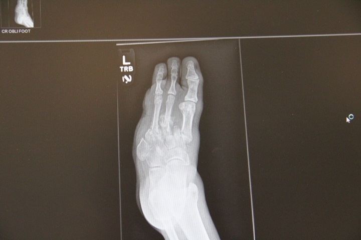
Bone cultures are recommended to guide antibiotic selection in cases of osteomyelitis. Plain radiographs should be taken of all diabetic foot ulcers to screen for retained foreign bodies, osteomyelitis, and subcutaneous gas.
X-ray findings of osteomyelitis take four to six weeks to develop, therefore, if there is a high index of suspicion of osteomyelitis, an MRI is helpful in making this diagnosis early. CT scans are useful for identifying deep space abscesses when MRI is not readily available.
Transcutaneous oxygen measurements (TCOM) can be helpful in risk stratifying these patients. Transcutaneous oxygen tension of at least 40 mmHg is needed for normal wound healing. Most patients with diabetes mellitus will have levels lower than this, and hyperbaric oxygen therapy and correction of peripheral vascular disease can increase oxygenation. Transcutaneous oxygen measurements can also be used to predict who will be most likely to benefit from hyperbaric oxygen therapy and who needs critical reperfusion. A transcutaneous oximetry measurement (TCOM) of greater than 200 mmHg under hyperbaric conditions indicates a greater likelihood of healing with a course of hyperbaric oxygen therapy with a sensitivity of 80% and positive predictive value of 88%[7].
Treatment / Management
Management strategies for wound care should include maintaining a moist wound environment, treating and preventing infection, offloading the affected area, debridement of necrotic tissue and biofilm, maximization of perfusion, nutrition and oxygen delivery. Offloading strategies include debridement of calluses, padding, orthotics, therapeutic footwear, walking boots, total contact casts, and Achille's tendon lengthening[8][9]. The presence of callous increases the likelihood of ulcer formation 11-fold and debriding the callous lowers the pressure exerted by 26%. The combination of Achille's tendon lengthening and total contact casts have the highest success of healing forefoot ulcers[10][11].
Superficial wound infections can be treated with topical antimicrobials, however, once cellulitis is present systemic antibiotics will be required. Even mild peripheral vascular disease should be corrected in these patients to optimize their likelihood of healing.
The duration of treatment with antibiotics usually varies from 2-4 weeks. Those with osteomyelitis may need at least 6 weeks of treatment. Hyperbaric oxygen therapy can prevent amputations in patients with Wagner grade 3 or higher ulcers with a number needed to treat of four[12].
Hyperbaric oxygen does this through a variety of mechanisms including increased oxygen delivery to ischemic/hypoxic tissues, enhanced white blood cell-mediated bacterial killing, angiogenesis, accelerated collagen synthesis and fibroblast growth, and decreased edema. Nine randomized controlled trials show the effectiveness of hyperbaric oxygen therapy in these patients in speeding healing, preventing amputations and improved transcutaneous oxygen measurements. Hyperbaric oxygen increases the likelihood of wound healing with an odds ratio of 10. A standard treatment course is 30 to 40 treatments at 2.4 ATA these are given once or twice daily[12].
Surgical debridement is an essential treatment in many diabetic foot infections. The surgery involves debridement of the infected bone. In some cases, digital amputation may be required. A vascular surgeon should be consulted prior to undertaking any debridement because some patients may benefit from a bypass of the occluded vessel.
Differential Diagnosis
- Thrombophlebitis
- Phlegmasia cerulean dolens
- Compartment syndrome
- Acutely cold limb
- Venous stasis ulcer
Staging
Current guidelines regarding diabetic foot osteomyelitis
- Control the soft tissue infection before resecting bone. Drain all infected areas
- Use negative pressure wound therapy dressings in between surgeries to help with healing
- Resect diseased bone until healthy bone is visible
- Any infected area of bone seen on x-ray should be removed
- All bone fragments removed at surgery should be examined for infection and organisms
- Sample proximal bone to ensure it is infection-free
- Use a power saw to resect bone
- Delayed primary closure should take place once all infection is cured
- Consider adjunctive tendo-Achilles lengthening in patients with ankle equinus deformity
- If the patient has significant forefoot functional issues which may trigger ulcers, consult a podiatrist
Prognosis
The prognosis for a diabetic foot infection depends on many factors including vascular blood supply and the presence of neuropathy. In many studies, two common risk factors for amputation include renal failure and significant peripheral vascular disease. Over the past 2 decades, the presence of MRSA is also deemed to be a risk factor for the progression of the infection. For patients who fail to control their blood glucose, the outcomes are poor; eventually resulting in an amputation. Even after a bypass, cure is not guaranteed as the surgery is demanding and failures are common. The key reason for surgical bypass failures is that the target vessel is very small. Patients who do undergo a limb amputation are also at risk for adverse cardiac events and stroke. The overall quality of life of patients with diabetes mellitus who develop a foot infection is poor.
Complications
Osteomyelitis
Bone fracture
Sepsis
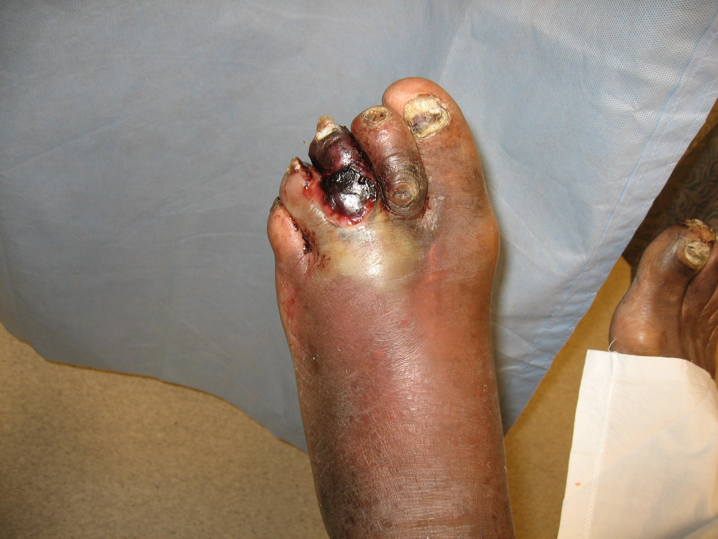
Gangrene
Necrosis
Amputation
Pearls and Other Issues
In summary, diabetic foot ulcers are precursors to amputations and require aggressive treatment. Patients should be screened for risk factors and referred for diabetic footwear when risk is identified. Staphylococcus aureus is the most important pathogen in diabetic foot infections, but as the depth and severity increase, these become polymicrobial. Surgery is indicated for the reversible peripheral vascular disease. Callouses should be debrided, and a moist wound healing environment should be maintained. Achille's tendon lengthening combined with total contact casting speeds healing and decreases the likelihood of recurrence and hyperbaric oxygen therapy is indicated in selected cases (Wagner grade 3 or higher) to speed healing and prevent amputation[13].
Enhancing Healthcare Team Outcomes
Diabetic foot ulcers have challenging and serious implications if not adequately managed. It is vital to follow an interprofessional approach when treating patients with diabetes mellitus who present with foot ulcers given their immunocompromised status and low threshold to fight superimposed infections and capacity to heal adequately. Comprehensive history and examination is essential in order to adequately formulate a treatment plan for patients with Diabetic foot ulcers. They should be managed by a team of healthcare professionals including Primary medical doctor, endocrinologist, podiatry, vascular surgery, infectious disease specialist, wound care nurse and pharmacist. The team should work closely in achieving optimal glycemic control and titrating diabetic regimen along with an interprofessional approach for adequate debridement, proper local wound care, redistribution of pressure on the ulcer by mechanical offloading, and control of infection and ischemia. The following approach should be followed in the management of diabetic foot ulcers:
- Primary medical doctor/endocrinologist should assess the wound and adequate referral to Podiatry should be provided. Diabetic foot ulcer should be classified upon initial presentation and monitored for any signs of worsening on regular follow-ups.
- Vascular surgery should be consulted for any evidence of underlying PAD ( Peripheral arterial disease) and signs of limb ischemia. Early revascularization is the key. Revascularization should also be performed in patients with a nonhealing ulcer and any degree of limb ischemia.
- Surgical (sharp) debridement versus utilization of autolytic debridement with hydrogels by wound care specialist should be performed as warranted.
- Infectious disease specialist opionion should be sought for the management of wound infections.
- Regular wound care in a clinic and closely followed by a wound care nurse
- The pharmacist should encourage the patient to discontinue smoking and maintain strict compliance with diabetic medications
- The patient should be seen by a dietitian to ensure that he/she is eating a healthy diet and maintaining a healthy weight.
There is also clear evidence about the effectiveness of hyperbaric oxygen therapy in the patients with Wagner Grade 3 ulcers in speeding healing, preventing amputations and improved transcutaneous oxygen measurement. In order to avoid the morbidity of a diabetic foot infection, the entire team must communicate to ensure that the patient is receiving appropriate therapy and guidance. [12]( Level 1)