Bowel Obstruction

Earn CME/CE in your profession:


Continuing Education Activity

A bowel obstruction can either be a mechanical or functional obstruction of the small or large intestines. Obstruction frequently causes abdominal pain, nausea, vomiting, constipation, obstipation, and distention. This activity explains the pathophysiology, classification, evaluation, and management in patients with bowel obstruction. It highlights the role of the interprofessional team in treating and decreasing long term morbidity in patients with bowel obstruction.

Objectives:

  • Describe the pathophysiology, epidemiology, and classification of bowel obstruction.
  • Outline the evaluation in a patient with bowel obstruction.
  • Explain the conservative and surgical management strategies in patients with bowel obstruction.
  • Explain the importance of a cohesive, interprofessional team approach to caring for patients with bowel obstruction.

Introduction

A bowel obstruction can either be a mechanical or functional obstruction of the small or large intestines. The obstruction occurs when the lumen of the bowel becomes either partially or completely blocked. Obstruction frequently causes abdominal pain, nausea, vomiting, constipation-to-obstipation, and distention. This, in turn, prevents the normal movement of digested products. Small bowel obstructions (SBOs) are more common than large bowel obstructions (LBOs) and are the most frequent indication for surgery on the small intestines. Bowel obstructions are classified as a partial, complete, or closed loop. A closed-loop obstruction refers to a type of obstruction in the small or large bowel in which there is complete obstruction distally and proximally in the given segment of the intestine.[1][2][3]

Etiology

There are many potential etiologies of small and large bowel obstructions that are classified as either extrinsic, intrinsic, or intraluminal. The most common cause of SBOs in industrialized nations is from extrinsic sources, with post-surgical adhesions being the most common. Significant adhesions can cause kinking of the bowel leading to obstruction. It is estimated that at least two-thirds of patients with previous abdominal surgery have adhesions. Other common extrinsic sources include cancer, which causes compression of the small bowel leading to obstruction. Less common but still prevalent extrinsic causes are inguinal and umbilical hernias. Untreated or symptomatic hernias may eventually become kinked as the small bowel protrudes through the defect in the abdominal wall and becomes entrapped in the hernia sack. Hernias that are not identified or are not reducible may progress to obstruction of the bowel and are considered a surgical emergency with the strangulated or incarcerated bowel becoming ischemic over time. Other causes of SBO include intrinsic disease, which can create an insidious onset of bowel wall thickening. The bowel wall slowly becomes compromised, forming a stricture. Crohn disease is the most common cause of benign stricture seen in the adult population.  [4][5]

Intraluminal causes for SBOs are less common. This process occurs when there is an ingested foreign body that causes impaction within the lumen of the bowel or navigates to the ileocecal valve and is unable to pass, forming a barrier to the large intestine. However, it is noted that most foreign bodies that pass through the pyloric sphincter will be able to pass through the rest of the gastrointestinal tract. LBOs are less common and compromise only 10% to 15% of all intestinal obstructions. The most common cause of all LBOs is adenocarcinoma, followed by diverticulitis and volvulus. Colonic obstruction is most commonly seen in the sigmoid colon.     

Epidemiology

Small and large bowel obstructions are similar in incidence in both males and females. The overriding factor affecting incidence and distribution depends on patient risk factors, including but not limited to: prior abdominal surgery, colon or metastatic cancer, chronic intestinal inflammatory disease, existing abdominal wall and/or an inguinal hernia, previous irradiation, and foreign body ingestion. [6][7]

Pathophysiology

The normal physiology of the small intestine consists of the digestion of food and the absorption of nutrients. The large bowel continues to aid in digestion and is responsible for vitamin synthesis, water absorption, and bilirubin breakdown. Any obstructive mechanism will hinder these physiologic components. Obstruction causes dilation of the bowel proximal to the transition point and collapses distally. A result of partial or complete blockage of digested products during obstruction is emesis. Frequent emesis can lead to fluid deficits and electrolyte abnormalities. As the condition is left untreated and worsens, a bowel wall edema forms, and third-spacing begins. A serious and life-threatening complication of bowel obstruction is strangulation. Strangulation is more commonly seen in closed-loop obstructions. If the strangulated bowel is not treated promptly, it eventually becomes ischemic, and tissue infarction occurs. Tissue infarction progresses to bowel necrosis, perforation, and sepsis/septic shock. 

History and Physical

Suspected bowel obstruction requires the practitioner to obtain a detailed medical history inquiring about significant risk factors related to bowel obstruction. Small and large bowel obstruction have many overlapping symptoms. However, quality, timing, and presentation differ. Commonly in SBO, abdominal pain is described as intermittent and colicky but improves with vomiting, while the pain associated with LBO is continuous. The vomiting in SBO tends to be more frequent, in larger volumes, and bilious, which is in contrast to vomiting during an LBO, which typically presents as intermittent and feculent when present. Tenderness to palpation is present in both conditions, but with SBO, it is more focal, and with LBO, it is more diffuse.

Additionally, distention is marked in LBO with obstipation more commonly present. It is important to note that in certain situations, an LBO will mimic an SBO if the ileocecal valve is incompetent. An incompetent ileocecal valve can allow for the insufflation of air from the large bowel into the small bowel producing symptoms of an SBO.

Evaluation

Although bowel obstruction alone can be suspected with an accurate patient history and presentation, the current standard of care to confirm the diagnosis in small and large bowel obstruction is an abdominal CT with oral contrast. CT allows for visualization of the transition point, the severity of obstruction, potential etiology, and assessment of any life-threatening complications. This information enables the provider to be more effective in identifying patients who will require surgical intervention.  Laboratory evaluation is essential to evaluate for any leukocytosis, electrolyte derangements that may be present as a result of the emesis. Labs also evaluate for elevated lactic acid that may be suggestive of sepsis or perforation, which at times may not be visible on CT if it is a microperforation and early in the course, blood cultures, or other signs of sepsis/septic shock. Although the lactic acid is often looked to in order to determine if there is a sign of perforation or ischemic gut, it should be noted this can be normal even with a microperforation present, initially. Physical examination of the patient remains an essential diagnostic tool regarding the patient's severity and the need for emergent surgery vs. medical management.[8]

Treatment / Management

Initial management should always include an assessment of the patient's airway, breathing, and circulation. If resuscitation is required, it should be performed with isotonic saline and electrolyte replacement. A Foley catheter should be inserted to monitor the patient's urine output if the patient is unstable or septic. Nasogastric tube insertion will allow for bowel decompression to relieve distention proximal to the obstruction. Nasogastric tube insertion will also help control emesis, allow for accurate assessment of intake and output, and lower the risk of aspiration.

Management ultimately depends on the etiology and severity of the obstruction. Stable patients with partial or low-grade obstruction resolve with nasogastric tube decompression and supportive measures. Patients who present with reducible hernias will require non-emergent surgical intervention to prevent future recurrence. Non-reducible or strangulated hernias require emergency surgical intervention. Complete or high-grade obstructions often require urgent or emergent surgical intervention as the risk of ischemia increases. Chronic disease states such as Crohn disease and malignancy require initial supportive measures and longer periods of nonoperative management. Treatment will ultimately depend on the patient's disposition and surgeon's acumen.

Differential Diagnosis

  • Abdominal hernias
  • Abdominal pain in elderly people
  • Appendicitis
  • Chronic megacolon
  • Colonic polyps
  • Diverticulitis
  • Diverticulitis empiric therapy
  • Pseudomembranous colitis surgery
  • Small bowel obstruction
  • Toxic megacolon

Prognosis

When bowel obstruction is managed promptly, the outcome is good. In general, when bowel obstruction is managed non surgically the recurrence rate is much higher than those treated surgically.

Complications

  • Intraabdominal abscess
  • Sepsis
  • Disability
  • Wound dehiscence
  • Aspiration
  • Short bowel syndrome
  • Pneumonia
  • Bowel perforation
  • Respiratory failure
  • Anastomotic leak
  • Renal failure
  • Death

Postoperative and Rehabilitation Care

The postoperative recovery, in most cases of bowel obstruction, is slow. These patients need prophylaxis against deep venous thrombosis and prevention of atelectasis. Ambulation is necessary. Time to feeding can vary depending on the ileus.

Consultations

  • General surgeon
  • Radiologist for drainage of any abscess
  • Stoma nurse
  • Infectious disease

Pearls and Other Issues

Most bowel obstructions will require hospital admission and surgical consultation. Prompt recognition and diagnosis are critical in improving morbidity and mortality. The most important step in the initial management of bowel obstruction is identifying the type, severity, and cause. Understanding the difference between emergent and non-emergent surgical intervention is essential in improving outcomes and preventing sequelae of complications, including bowel necrosis, perforation, and sepsis. Disposition ultimately depends on the type and etiology of the obstruction, as well as the patient's past medical history, current health status, and risk factors.

Enhancing Healthcare Team Outcomes

The key to preventing the high mortality following a bowel obstruction is the early diagnosis, resuscitation, and operative intervention. An interprofessional team is vital to ensure that the patient receives prompt attention. The triage nurse must be fully aware of the signs of bowel obstruction and expedite the admission. The emergency physician, nurse practitioner, or physician assistant must examine the patient and get the appropriate radiological test. The surgeon must be consulted even if no intervention is planned. While awaiting surgery, the bowel may need to be decompressed with a nasogastric tube, and the nurse is essential for monitoring of vital signs and worsening of the obstruction. Communication between healthcare workers is critical. [9][4] [Level V]

Outcomes

The morbidity and mortality of bowel obstruction are dependent on early diagnosis and management. If any strangulated bowel is left untreated, there is a mortality rate of close to 100%. However, if surgery is undertaken within 24-48 hours, the mortality rates are less than 10%. Factors that determine the morbidity include the age of patient, comorbidity, and delay in treatment. Today, the overall mortality of bowel obstruction is still about 5%-8%.[3][10] [Level 3]


<p>Contributed by Michael Schick DO, MA.</p>

(Click Image to Enlarge)
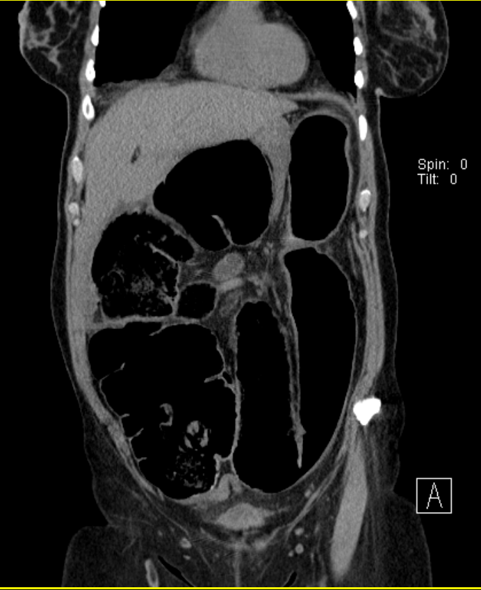
FIGURE 5: Coronal CT abdomen reveals cecal volvulus
FIGURE 5: Coronal CT abdomen reveals cecal volvulus. Usually a patient with a cecal volvulus will present with small and large bowel obstructions, with collapse of the distal large bowel, and with extensive dilation of the proximal small bowel.
Contributed by Wyckoff Height Medical Center 2017

(Click Image to Enlarge)
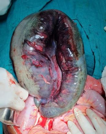
Sigmoid vulvulus
Sigmoid vulvulus
Contributed by Sunil Munakomi, MD

(Click Image to Enlarge)
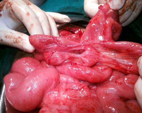
adhesive intestinal obstruction
Contributed by Sunil Munakomi, MD
Details

Editor:

Sara M. Nehring

Updated:

7/31/2023 8:39:17 PM

References


[1]

van Steensel S, van den Hil LCL, Schreinemacher MHF, Ten Broek RPG, van Goor H, Bouvy ND. Adhesion awareness in 2016: An update of the national survey of surgeons. PloS one. 2018:13(8):e0202418. doi: 10.1371/journal.pone.0202418. Epub 2018 Aug 17     [PubMed PMID: 30118503]

Level 3 (low-level) evidence

[2]

Behman R, Nathens AB, Karanicolas PJ. Laparoscopic Surgery for Small Bowel Obstruction: Is It Safe? Advances in surgery. 2018 Sep:52(1):15-27. doi: 10.1016/j.yasu.2018.03.001. Epub 2018 Jun 19     [PubMed PMID: 30098610]

Level 3 (low-level) evidence

[3]

Behman R, Nathens AB, Look Hong N, Pechlivanoglou P, Karanicolas PJ. Evolving Management Strategies in Patients with Adhesive Small Bowel Obstruction: a Population-Based Analysis. Journal of gastrointestinal surgery : official journal of the Society for Surgery of the Alimentary Tract. 2018 Dec:22(12):2133-2141. doi: 10.1007/s11605-018-3881-z. Epub 2018 Jul 26     [PubMed PMID: 30051307]


[4]

Ten Broek RPG, Krielen P, Di Saverio S, Coccolini F, Biffl WL, Ansaloni L, Velmahos GC, Sartelli M, Fraga GP, Kelly MD, Moore FA, Peitzman AB, Leppaniemi A, Moore EE, Jeekel J, Kluger Y, Sugrue M, Balogh ZJ, Bendinelli C, Civil I, Coimbra R, De Moya M, Ferrada P, Inaba K, Ivatury R, Latifi R, Kashuk JL, Kirkpatrick AW, Maier R, Rizoli S, Sakakushev B, Scalea T, Søreide K, Weber D, Wani I, Abu-Zidan FM, De'Angelis N, Piscioneri F, Galante JM, Catena F, van Goor H. Bologna guidelines for diagnosis and management of adhesive small bowel obstruction (ASBO): 2017 update of the evidence-based guidelines from the world society of emergency surgery ASBO working group. World journal of emergency surgery : WJES. 2018:13():24. doi: 10.1186/s13017-018-0185-2. Epub 2018 Jun 19     [PubMed PMID: 29946347]

Level 1 (high-level) evidence

[5]

Pavlidis E, Kosmidis C, Sapalidis K, Tsakalidis A, Giannakidis D, Rafailidis V, Koimtzis G, Kesisoglou I. Small bowel obstruction as a result of an obturator hernia: a rare cause and a challenging diagnosis. Journal of surgical case reports. 2018 Jul:2018(7):rjy161. doi: 10.1093/jscr/rjy161. Epub 2018 Jul 3     [PubMed PMID: 29992011]

Level 3 (low-level) evidence

[6]

Andersen P, Jensen KK, Erichsen R, Frøslev T, Krarup PM, Madsen MR, Laurberg S, Iversen LH. Nationwide population-based cohort study to assess risk of surgery for adhesive small bowel obstruction following open or laparoscopic rectal cancer resection. BJS open. 2017 Apr:1(2):30-38. doi: 10.1002/bjs5.5. Epub 2017 Jul 26     [PubMed PMID: 29951603]


[7]

Doshi R, Desai J, Shah Y, Decter D, Doshi S. Incidence, features, in-hospital outcomes and predictors of in-hospital mortality associated with toxic megacolon hospitalizations in the United States. Internal and emergency medicine. 2018 Sep:13(6):881-887. doi: 10.1007/s11739-018-1889-8. Epub 2018 Jun 12     [PubMed PMID: 29948833]


[8]

Li PH, Tee YS, Fu CY, Liao CH, Wang SY, Hsu YP, Yeh CN, Wu EH. The Role of Noncontrast CT in the Evaluation of Surgical Abdomen Patients. The American surgeon. 2018 Jun 1:84(6):1015-1021     [PubMed PMID: 29981641]


[9]

Pisano M, Zorcolo L, Merli C, Cimbanassi S, Poiasina E, Ceresoli M, Agresta F, Allievi N, Bellanova G, Coccolini F, Coy C, Fugazzola P, Martinez CA, Montori G, Paolillo C, Penachim TJ, Pereira B, Reis T, Restivo A, Rezende-Neto J, Sartelli M, Valentino M, Abu-Zidan FM, Ashkenazi I, Bala M, Chiara O, De' Angelis N, Deidda S, De Simone B, Di Saverio S, Finotti E, Kenji I, Moore E, Wexner S, Biffl W, Coimbra R, Guttadauro A, Leppäniemi A, Maier R, Magnone S, Mefire AC, Peitzmann A, Sakakushev B, Sugrue M, Viale P, Weber D, Kashuk J, Fraga GP, Kluger I, Catena F, Ansaloni L. 2017 WSES guidelines on colon and rectal cancer emergencies: obstruction and perforation. World journal of emergency surgery : WJES. 2018:13():36. doi: 10.1186/s13017-018-0192-3. Epub 2018 Aug 13     [PubMed PMID: 30123315]


[10]

Mellor K, Hind D, Lee MJ. A systematic review of outcomes reported in small bowel obstruction research. The Journal of surgical research. 2018 Sep:229():41-50. doi: 10.1016/j.jss.2018.03.044. Epub 2018 Apr 16     [PubMed PMID: 29937015]

Level 1 (high-level) evidence