Introduction
Our somatosensory system has three basic types of sensory receptors that detect different types of external stimuli. These include mechanoreceptors that detect light touch, vibration, pressure, and texture; nociceptors that detect pain; and thermoreceptors that detect temperature. Mechanoreceptors further divide into Merkel disks, Meissner’s corpuscles, Ruffini endings, and Pacinian corpuscles based on the specific type of mechanical stimuli they perceive. The perception of vibratory sensation is by two main types of mechanoreceptors, Meissner corpuscles (MC) and Pacinian corpuscles (PC). MCs are large myelinated fibers that detect low-frequency vibration and are present in glabrous (smooth, hairless) skin on fingertips and eyelids. PCs contain large myelinated A-beta fibers which are rapidly adapting and are present in the deeper layers of skin, ligaments, and joints where their function is to detect high-frequency vibration and deep pressure.
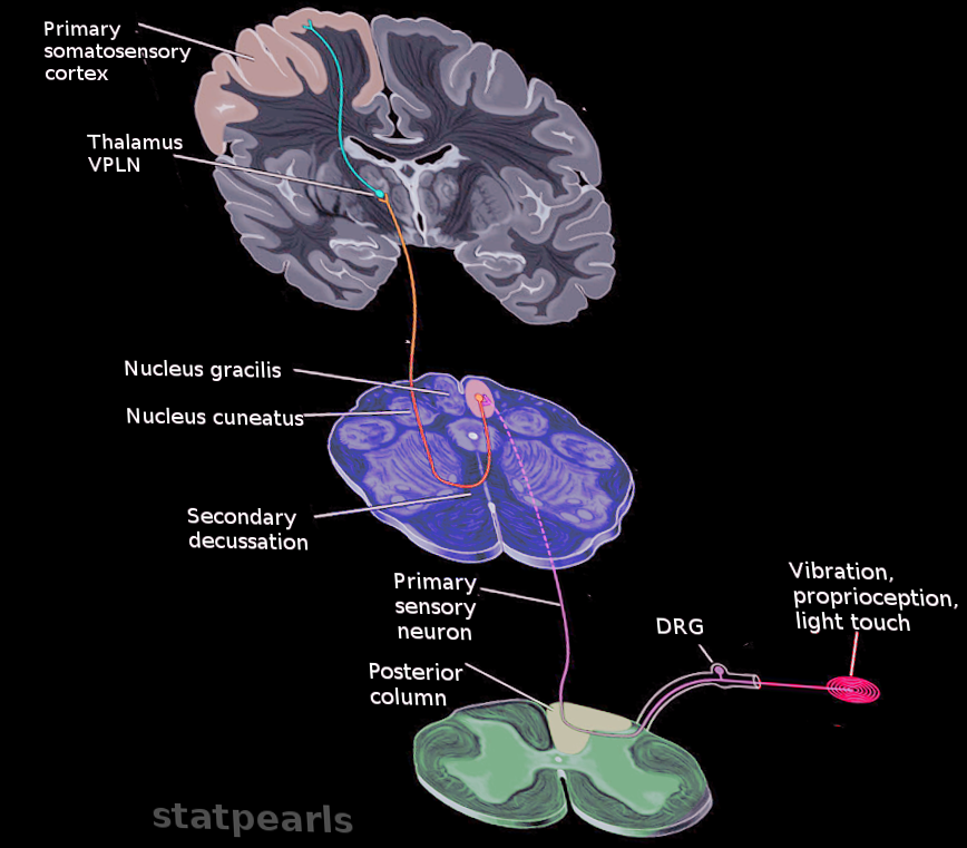
The vibratory sensations transmit via the dorsal column of the spinal cord to the primary somatosensory area of cortex. The sensory signals from Meissner and Pacinian corpuscles terminate in the cell body of dorsal root ganglion (pseudounipolar neurons) and constitute the first-order neuron of the dorsal column. The axonal processes leave the dorsal horn gray matter and enter the dorsal funiculus to constitute either the fasciculus gracilis or the fasciculus cuneatus. Fasciculus gracilis carries sensory information from the lower limbs and synapses with the nucleus gracilis in the caudal medulla. It is located medially in the dorsal column as compared to fasciculus cuneatus which is located laterally and carries sensory information from the upper extremities and synapses with nucleus cuneatus in caudal medulla. That is why fasciculus cuneatus is only present at spinal level T6 and above. Nucleus gracilis and nucleus cuneatus constitute the second-order neurons of this pathway. The fibers from both sides decussate in caudal medulla as internal arcuate fibers and ascend contralaterally as medial lemniscus. The second synapse occurs in the ventral posterolateral nucleus (VPL) of the thalamus, which constitutes the third-order neurons of the pathway. Axons from VPL nucleus travel through the posterior limb of the internal capsule and terminate in the primary somatosensory cortex, located in the postcentral gyrus.[1][2][3]
Issues of Concern
The most significant concern related to vibration sensation is the peripheral neuropathy of diabetes mellitus, which affects the peripheral nerves and results in the loss of pain, temperature, and vibration sensations. Better glycemic control is the only way to prevent this pathology.[4]
Cellular Level
Pacinian corpuscles (PCs) are highly specialized corpuscles found in the dermis of glabrous skin. This specialization is partly due to the adventitial tissue surrounding the corpuscles. PCs have a large, ellipsoid shape, ranging up to 3 to 4mm in the digits. Their shape and size vary depending upon the location in the body and area of skin of their residence. PCs have a large myelinated centrally located nerve fiber called neurite, running along their long axis. Bilaterally arranged glial lamellae immediately surround the neurites, and perineural (collagenous) lamellae further surround them on the outer aspect. Inner core cells are derived from Schwann cells and are involved in the conduction of signals. These lamellar layers are separated from each other by a fluid-filled layer, kept in between due to the presence of tight junctions found between adjacent lamellar cells. A capsule surrounds the outer lamellar layer. When a vibratory stimulus deforms skin, this stimulus reaches the dermis where it transmits to the external capsule of PCs. This distortion travels through the external and internal core until it reaches the central neurite. As a result, stretch-gated cation channels (voltage-gated and non-voltage gated) on the neuronal membrane open and conduct a neural response.[5]
Development
The development of Pacinian corpuscles (PCs) starts at the gestational age of 13 weeks and is completed at the age of 4 months postnatal. The inner core forms from a compact network of Schwann cells and outer core, and the surrounding mesenchyme forms the capsule. The development of Meissner corpuscles (MCs) starts at 22 weeks and attains complete morphology by 8 months of life. The developing MCs have a complex relationship of their axons with the epidermis and are covered by Schwann cells until late maturity. The present data suggest that there is an asynchronous relationship between the development and immunohistochemical maturation of MCs and PCs.[6] The alar and basal plates of neural tube give origin to dorsal (sensory) and ventral (motor) horns of the spinal cord, respectively. The dorsal horn makes the sensory pathway of the posterior column, which carries the vibratory signals.
Organ Systems Involved
The sense of vibration is perceived and conducted to the brain via integumentary and nervous systems. Skin is involved in the conduction of distortion from the environment to the external capsule of PCs in the dermis, and from the central neurite of PCs, the neuronal signal transmits via the dorsal column-medial lemniscus to the VPL nucleus of the thalamus. From the VPL nucleus, the final transmission is to the somatosensory area of the cerebral cortex.[7]
Function
The function of vibratory receptors is to make us aware of different frequencies of external stimuli. Meissner corpuscles detect frequencies between 30 and 50 Hz; Pacinian corpuscles are sensitive to frequencies between 100 and 400 Hz. The dorsal column-medial lemniscus pathway appreciates fine touch, 2-point discrimination, and vibration sensations from all the body excluding the head.
Mechanism
Pacinian and Meissner corpuscles are located in the deep dermis and detect distortion of skin from external stimuli and transmit it to the central neurite. Stretch gated ion channels open and conduct a neural response; this is the underlying mechanism of vibration perception. Trigeminal nerve transmits the vibrotactile signals from the facial area, and the dorsal column–medial lemniscus (DCML) pathway carries vibratory signals from the rest of the body. The signals get relayed via the thalamus to the primary somatosensory area of the postcentral gyrus.[8][1]
Related Testing
The Romberg test assesses the integrity of the posterior column pathway, which carries vibratory sensations. The examiner asks the patient to close his/her eyes while standing. If the patient can not maintain his/her balance with eyes closed, the problem lies in the posterior column, and not in the cerebellum.
Pathophysiology
The pathophysiology of vibration perception and its pathways is related to the diseases affecting the dorsal column. Any factor which disrupts the neuronal transmission from vibratory sensors in the deep dermis to the somatosensory area in the cortex affects the vibratory sensations. The explanation of several of these mechanisms is in the clinical significance section below.
Clinical Significance
The clinical significance of vibratory sensation lies in its carrier pathway (DCML), which also carries position sense and fine touch. Several diseases affect the DCML pathway and thus manifest as loss of position, vibration, and touch sensations.
Most common in developed countries is subacute combined degeneration of the spinal cord due to vitamin B12 deficiency. Vitamin B12 primarily exists in animal products, including meat, fish, poultry, and milk. Our bodies have extensive reserves of B12 and symptoms of deficiency appear after several years of a diet devoid of B12, explaining why vegetarians are mostly the victims of vitamin B12 deficiency if they don’t substitute it with other sources.
Another example is tabes dorsalis, a late manifestation of tertiary syphilis. It causes degenerative joints disease (Charcot joints), Argyll Robertson pupils (light reflex lost but accommodation reflex retained), and affects dorsal column. Tabes dorsalis incidence is reduced dramatically after the introduction of penicillin in the treatment of syphilis.
An infarction of the posterior spinal artery also affects the posterior column pathway resulting in the loss of vibration, proprioception, and light touch below the level of the lesion.
Hemisection of the spinal cord, known as Brown-Sequard syndrome, also affects the dorsal column along with other pathways. It is commonly the result of trauma, tumors, and abscesses.[9][10][11]