Continuing Education Activity
Foot dislocations are uncommon injuries that can occur at multiple different joints of the foot. The three most common joints involved include the subtalar joint, the midtarsal joint, and the tarsometatarsal (TMT) joint complex. This activity outlines and reviews the evaluation and management of foot dislocations, and reviews the role of the healthcare team in improving care for patients with this condition.
Objectives:
- Identify the epidemiology of foot dislocations.
- Outline the appropriate evaluation of patients presenting with foot dislocations.
- Review the treatment and management options available for patients with foot dislocations.
- Describe interprofessional team strategies for improving care coordination and communication to advance the diagnosis and treatment of foot dislocations and improve outcomes.
Introduction
Foot dislocations are uncommon injuries that can occur at multiple different joints of the foot. This article will cover the three most commonly dislocated joints that make up the foot. These injuries can be pure dislocations or combined fracture-dislocations. The three most common joints involved include the subtalar joint, the midtarsal joint (Chopart joint), and the tarsometatarsal (TMT) joint complex, otherwise known as the “Lisfranc Joint.” Recognition of these injuries is important as subtle dislocations are commonly missed, especially Lisfranc injuries, on initial presentation, and can lead to osteoarthritis and long term disability if not treated correctly.[1]
Etiology
Just like in other joints of the body, specific mechanisms are more associated with certain foot dislocations. Dislocations of the foot can occur after either direct or indirect trauma. Common mechanisms include falling from a tall height, motor vehicle collisions, or sports injuries. Lisfranc injuries commonly occur following any of the above-stated mechanisms through indirect rotational forces and axial loading on a foot that is hyper-plantarflexed. Subtalar dislocations usually occur following a high energy mechanism such as a fall from heights or a high-speed motor vehicle collision (MVC) with the foot in either inversion or eversion. Midtarsal dislocations are also due to high energy mechanisms and occur when such forces are directed at a plantarflexed foot.
Epidemiology
Subtalar dislocations of the talus and calcaneus are the more common of the foot dislocations but are also very rare overall, accounting for around 1% of all dislocations. They are more common in young or middle-aged males. Midtarsal dislocations are the second most common fracture-dislocation involving the midfoot following those involving the Lisfranc joint.[2] Lisfranc injuries fracture-dislocations are the least common of the foot dislocations and account for around 0.2% of all fractures. They are seen more commonly in males in the third decade.
Pathophysiology
The foot consists of multiple bones and articulations. Overall, there are 26 bones, 33 articulations, and 3 functional and anatomic regions. The hindfoot consists of the talus and calcaneus, while the midfoot consists of the navicular, the cuboid, and the 3 cuneiforms. The Lisfranc joint, or forefoot, consists of 5 metatarsals and 14 phalanges. The articulations between the hindfoot and the midfoot make up the midtarsal or Chopart joints. They are made up of the talonavicular and the calcaneocuboid joints. The articulations between the midfoot and the forefoot are termed the Lisfranc joints and consist of 5 tarsometatarsal joints. These three articulations are the tarsometatarsal articulation, intermetatarsal articulation, and the intertarsal articulations. Flexion and extension primarily occur at the metatarsophalangeal (MTP) and interphalangeal (IP) joints.
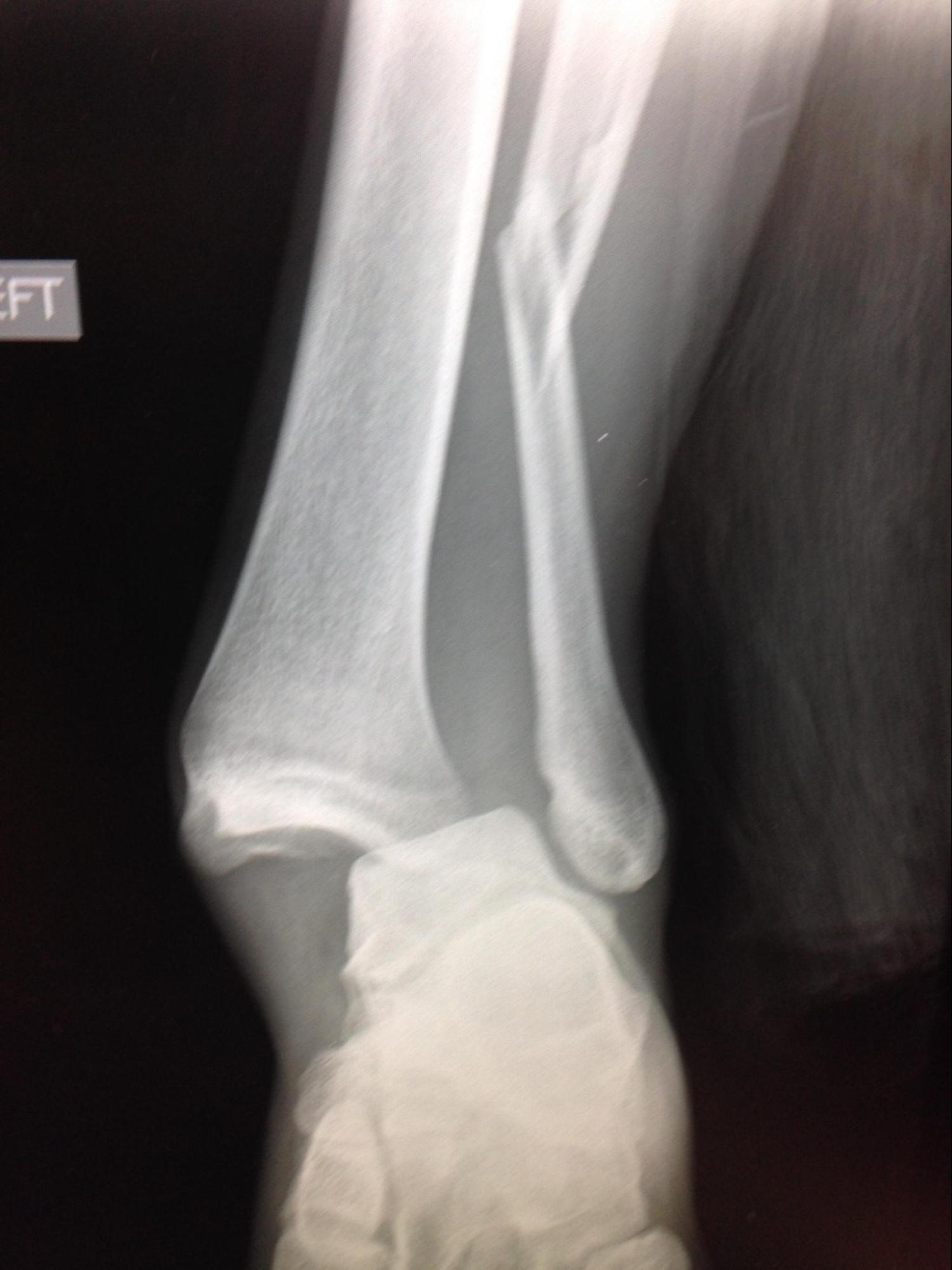
Subtalar / Peritalar Dislocation
A subtalar dislocation occurs when both the subtalar (talocalcaneal) and talonavicular dislocate. [3] Such dislocations are rare and often the result of vigorous trauma sustained from motor vehicle accidents or a fall from a significant height. However, several cases describe subtalar dislocations in basketball players after who perform repetitive jumping and landing. The considerable strength of the ligaments connecting the talus and calcaneus contributes to the rarity of subtalar dislocations. Subtalar dislocations are classified based on the direction of the dislocation of the foot relative to the talus. Medial dislocations are the most common type and are seen following inversion injuries, leaving the foot fixed in supination. Lateral dislocations are the second most common type and are due to eversion injuries, which cause the foot to be fixed in pronation. Up to 25% of these dislocations are open, especially if it is a lateral dislocation. Also, subtalar dislocations often occur with tarsal or metatarsal fractures.[3][4][5]
Midtarsal (Chopart) Joint Dislocation
Chopart joint dislocations are common injuries seen following substantial trauma to the midfoot area of an inverted ankle.[2] As with other foot dislocations, this trauma is usually in the form of motor vehicle collisions or falls from considerable heights. The axial forces sustained in such impacts typically leads to dislocation of the navicular bone from the talus, with possible concomitant fractures of the talus, navicular, cuboid, and calcaneus. The dislocation of the calcaneocuboid joint leads to less displacement than that seen with the talonavicular dislocation, and but can lead to comminuted fractures of the cuboid and calcaneus.[2]
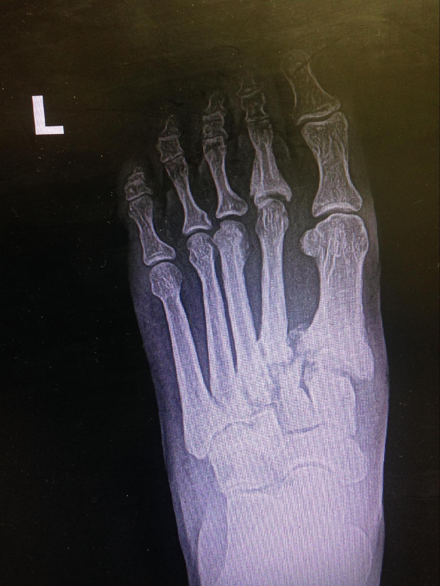
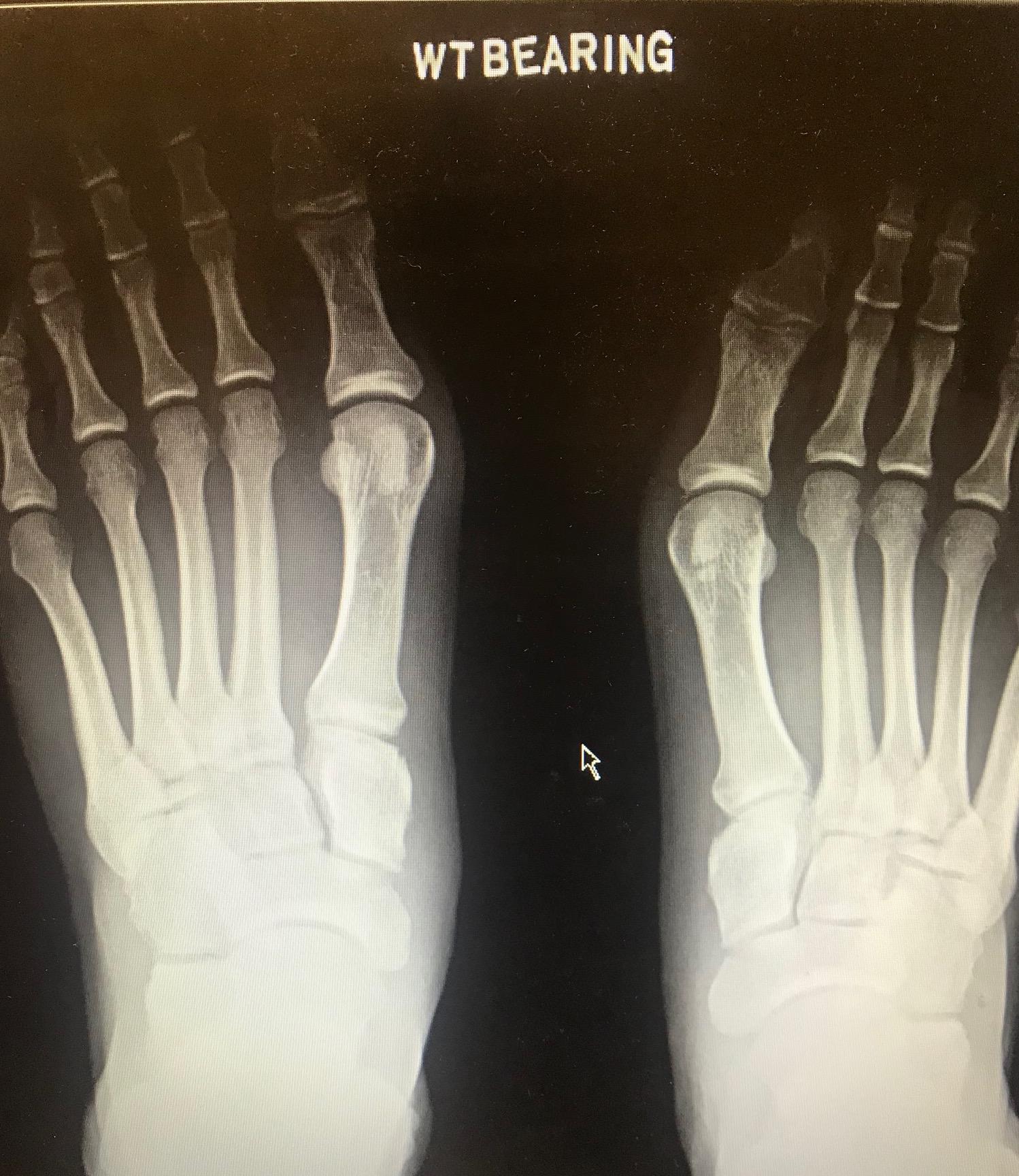
Lisfranc/Tarsometatarsal Dislocation
The Lisfranc/tarsometatarsal joint is comprised of the articulations between the first 3 metatarsals and the 3 cuneiform bones: medial, intermediate, and lateral.[6] Thus, a Lisfranc dislocation is a broad term describing any dislocation involving these many articulations. Lisfranc dislocations are due to either direct or indirect mechanisms. Indirect injuries are the more common type and are typically seen in athletes as the result of a longitudinal and medial/lateral rotational force directed at a plantarflexed foot. The forefoot is forced into hyperplantarflexion, which leads to tearing of the dorsal ligaments. These types of injuries typically lead to solely ligamentous damage, but smaller avulsion-type fractures of the metatarsals are also possible. Direct injuries involve crushing forces to the joint, usually seen in motor vehicle collisions or industrial accidents. These injuries are associated with more traumatic bony injuries than those seen in indirect injuries.[6][7]
Total Talar Dislocation
A total talar dislocation is a rare form of foot dislocation where the talus dislocates from all three of its articulations: the talonavicular, talocalcaneal, and tibiotalar joints. Such dislocation is usually the result of substantial trauma to an inverted and plantar-flexed foot, often in the form of a motor vehicle collision or falls from a substantial height.[8] Approximately half of all total talar dislocations are associated with fractures, most often the talus, and nearly all cases are associated with an open injury.[9] Large open wounds at the anterolateral ankle can be found with extruded talus. The talus should be preserved for reimplantation.
History and Physical
It is essential to obtain a thorough history and mechanism of injury when evaluating a patient with a foot injury. The mechanism alone can help clue the clinician into potential injuries of the foot. Patients will often not be able to weight-bear following the injury, which will help clue the examiner in on a possible dislocation or fracture being present.
On physical exam, one of the first things an examiner will notice upon the inspection are signs of bruising or if an obvious deformity is present. Lisfranc injuries typically present with a swelling in the midfoot, a foot locked in plantar flexion, and findings of ecchymosis on the plantar surface are pathognomonic.[6] Patients with Lisfranc injuries also report mid-foot pain with weight-bearing, particularly when walking downstairs. Provocative tests suggesting Lisfranc injury include pain upon palpation of the midfoot as well as the following specific tests:
- The piano key test, where the provider grasps the metatarsals of the affected foot and performs plantarflexion and dorsiflexion at the tarsometatarsal joint, noting any pain or subluxation.
- Compression of the midfoot with plantar flexion and dorsiflexion of the first metatarsal head relative to the second
- Compression along the wide-axis of the foot to induce stress between the first and second metatarsals, noting any pain or palpable clicks
- Stabilization of the hindfoot while the forefoot is passively pronated and abducted, noting any pain.
Visual clues for subtalar dislocation are dramatic and usually present. When a medial subtalar dislocation, the foot will be locked in supination, “acquired clubfoot.” When a lateral dislocation is present, the foot will be locked in pronation, “acquired flatfoot.” Lisfranc dislocations normally present with the foot locked in plantarflexion.
After inspection, palpation can clue the examiner in on swelling and tenderness of focal areas of the foot that is injured. Additionally, it is important to palpate and check for potential vascular compromise or any neurological injuries. The clinician performs this through the evaluation of the patient’s dorsalis pedis (DP) and posterior tibial (PT) pulses. Capillary refill should also be examined and documented before and following closed reduction.
Evaluation
Multiple imaging modalities can be used to demonstrate dislocations. Conventional radiography can be used to identify frank diastasis at the tarsometatarsal (TMT) joint. Computed tomography (CT) can also detect minimal subluxations and Lisfranc injuries. Ultrasonography can be used to identify dorsal Lisfranc ligament (DLL) length and thickness. However, the best imaging modality for ligament abnormalities is magnetic resonance imaging (MRI).[10] High-energy injuries often can be diagnosed with non-weight-bearing imaging. Low-energy injuries may require additional weight-bearing imaging if initial radiographs are ambiguous; these images become more important when evaluating for possible Lisfranc injuries.[11] Subtalar dislocations are easy to spot clinically as the deformation is clear, and standard X-rays displaying movement of the talus into the tibiofibular mortise confirm the diagnosis.[12] Midtarsal dislocations are initially evaluated using AP, lateral, and oblique x-rays of the foot and ankle. The talonavicular joint is best profiled on the AP view of the foot and the lateral view of the ankle or foot while the calcaneocuboid joint can be best seen on the medio-plantar oblique view of the foot.[2] If x-rays are not sufficient, then CT scans might be required to see small fractures or to aid in operative planning. The osseous trauma sustained in such fracture-dislocations takes precedence over any soft tissue injuries, and thus MRI is often not utilized in the initial evaluation.[2]
Treatment / Management
An accurate diagnosis is paramount in the management of foot injuries. It is important to first properly identify which dislocation has occurred to formulate a treatment plan. Reduction of dislocation should be done immediately to reduce avascular necrosis risk. Minor sprains with no joint displacement can be treated conservatively with initial immobilization and avoidance of weight-bearing. Ligamentous injuries can be treated with partial arthrodesis.
When a subtalar fracture or dislocation is present, it can be treated with numerous different methods of surgical internal fixations and/or joint fusions. Early closed reduction with sedation is performed if possible, but an open reduction is necessary if there is soft tissue obstruction or locked dislocations. These operations have varying efficacy, with a common goal to improve overall foot anatomy and function and relieve pain.
Lisfranc injuries can have serious complications if left untreated. Immediate surgical intervention is often necessary. The gold standard treatment for Lisfranc injuries is surgical open reduction and internal fixation, accompanied by arthrodesis. A new and promising technique for ORIF of Lisfranc injuries employs a Lapidus plate system, which has shown success in several reported cases. Although few cases have been reported, less invasive surgical options for Lisfranc injuries such as percutaneous fixation have been noted as effective alternatives to open reduction and internal fixation in patients with certain types of Lisfranc injuries.[13][14]
In most cases of midtarsal dislocation, closed reduction is initially attempted; however, the majority of these injuries eventually require open reduction and fixation.[2] In cases of severe trauma, the fracture-dislocation be temporarily externally fixated before the definitive internal fixation once other more pressing issues have been addressed.[2]
If surgery is performed, reevaluation of the affected foot is necessary. A physical exam of the affected foot, as well as repeat imaging, should be done to determine the efficacy of the surgical intervention.
Initial pain control is usually achieved with intravenous opioids such as morphine and fentanyl along with intravenous ketorolac. Following acute management, oral medications such as NSAIDs, acetaminophen, and oral opioids can be used to limit the duration of use of opioids when possible.
The type of injury will dictate the functional limitations post-injury. As a general rule, patients suffering from any foot dislocation will usually be non-weight bearing following the injury whether or not surgery is required or not. Following surgery, this can be for up to six to eight weeks. After this period, a walking boot or cast will be used and worn for another amount of time, and this is solely determined by the type of injury and rehabilitation status of the patient.
Differential Diagnosis
The differential diagnosis of patients initially presenting with undifferentiated atraumatic foot pain include:
- Stress fracture
- Soft-tissue injury
- Ligamentous injury
- Plantar fasciitis
- Morton’s neuroma
- Tarsal tunnel syndrome
- Nerve impingement
The differential diagnosis of patients initially presenting with undifferentiated traumatic foot pain include:
- Subtalar - peritalar dislocation
- Midtarsal (Chopart) joint dislocation
- Lisfranc - tarsometatarsal dislocation
- Total talar dislocation
- Fractures of the hindfoot
- Fractures of the midfoot
- Fractures of the forefoot
- Ligamentous injury of the hindfoot
- Ligamentous injury of the midfoot
- Ligamentous injury of the forefoot
Complications
Acute complications can occur but are rare. Compartment syndrome is a possible complication, and if suspected, compartment pressures should be checked with a hand-held manometer device. Additionally, amputation is a rare complication that can occur if Lisfranc injuries are left untreated.
Long term complications following foot dislocations are common. Patients commonly note stiffness following these injuries, along with many showing degenerative changes on follow up radiographs. As with fractures, dislocations, or sprains elsewhere in the body, dislocation of the joints of the foot can hasten the onset of osteoarthritis.[2]
Consultations
Patients suffering any of the above-listed types of foot dislocations should be referred to an orthopedic surgeon to ensure proper management. Some injuries require more emergent consultation, while some other subtle injuries can be referred to as an outpatient with proper discharge precautions.
Deterrence and Patient Education
Patients should be educated on the complexity of these injuries and long term sequelae such as osteoarthritis that can occur following foot dislocations. Educating them is paramount to ensure proper follow-up, discharge compliance with rehabilitation, and post-surgical instructions.
Enhancing Healthcare Team Outcomes
Foot dislocations are best managed by an interprofessional team that also includes nurses, technicians, and therapists. The diagnosis of foot dislocations is not always as simple as the more subtle injuries can be missed, and it is on the clinicians to know the proper imaging studies to obtain. This type of caution is more often needed when potential injuries to the forefoot are present as the can present is a less subtle manner. Imaging needed includes an anteroposterior, 30-degree oblique, and lateral weight-bearing radiographs. Despite their difficulty, weight-bearing films are essential in diagnosis and management, especially in subtle cases. Both the patient, nurses, and radiography technician should be educated on the importance of these injuries, thus allowing the proper X-rays to be obtained. Following stabilization or reduction, a physical therapist may work with the patient to restore function and strength, reporting back to the orthopedist regarding progress. Orthopedic nurses will monitor treatment progress, and inform the clinicians of any setbacks or lack of progress. They can also answer patient questions and coordinate care with therapists. These interprofessional steps can result in improved patient outcomes. [Level 5]