Central Line

Earn CME/CE in your profession:


Continuing Education Activity

Central venous catheterization (CVC) is a procedure frequently required in acute or critical care resuscitation. Indications include patients with multiple, incompatible intravenous (IV) medications with limited peripheral access, or who are being treated with vasoactive or phlebosclerotic agents which may not be suitably cared for with a peripheral IV alone. Some central lines are also placed for temporary or permanent hemodialysis access; these dialysis catheters are significantly larger than traditional double, triple, or quadruple lumen catheters placed in the emergency department or intensive care unit setting. Central lines may also be placed to introduce Swan Ganz catheters to measure internal hemodynamics of the heart, or to introduce temporary transvenous pacemaker leads in the critically ill patient who has severe bradycardia or high-degree heart block; these are called introducer catheters. Most central lines are placed today via the Seldinger technique, which is safer than the previously used "cut-down" technique. This activity reviews the indications, contraindications, and technique involved in performing central line placement and highlights the role of the interprofessional team in the care of patients undergoing this procedure.

Objectives:

  • Identify the indications for central line placement.
  • Describe the recommended patient position during central line placement.
  • Review the complications associated with central line placement.
  • Outline a structured interprofessional team approach to provide effective care to and appropriate surveillance of patients undergoing central line placement.

Introduction

Central venous catheterization (CVC) is a procedure frequently required in acute or critical care resuscitation. Indications include patients with multiple, incompatible intravenous (IV) medications with limited peripheral access, or who are being treated with vasoactive or phlebosclerotic agents which may not be suitably cared for with a peripheral IV alone. Some central lines are also placed for temporary or permanent hemodialysis access; these dialysis catheters are significantly larger than traditional double, triple, or quadruple lumen catheters placed in the emergency department (ED) or intensive care unit (ICU) setting. Central lines may also be placed to introduce Swan Ganz catheters to measure internal hemodynamics of the heart, or to introduce temporary transvenous pacemaker leads in the critically ill patient who has severe bradycardia or high-degree heart block: these are called introducer catheters. Most central lines are placed today via the Seldinger technique (a safety enhancement over the previous "cut-down" technique), in which the chosen vein is cannulated with a needle, a guidewire is inserted to maintain a tract through the skin into the vein, and the catheter is then inserted over the wire into the vein before the wire is removed. This procedure is generally performed with ultrasound guidance unless an ultrasound machine is unavailable or there are other exigent circumstances, in which case a palpation guided approach can be used. Despite the general overall safety of this procedure, complications do occur. This activity focuses on the complications of line placement.[1][2][3]

Anatomy and Physiology

Central venous catheters are placed typically in one of 3 large central veins: the internal jugular vein (IJ), subclavian vein (SCL), or femoral vein. These large diameter central veins are located universally near a large artery. In the lateral neck, the IJ is located next to the carotid artery, with the vein lying lateral to the artery in most patients, deep to the sternocleidomastoid muscle. The subclavian vein lies next to the subclavian artery, and courses out of the axilla across the lateral upper chest where it quickly dives deep to the clavicle before forming a confluence with the internal jugular vein which then becomes the brachiocephalic vein. The right and left brachiocephalic veins then join together to form a common superior vena cava. The femoral vein lies in the femoral triangle of each thigh and maintains a medial relationship to the common femoral artery.[4] A common mnemonic to remember the anatomic relationship of the femoral triangle is 'NAVeL' from lateral to medial indicating femoral nerve, femoral artery, femoral vein and lymphatics. Proximity to adjacent vital structures (arteries and lungs principally) is perhaps of greatest concern when striving to perform this procedure without causing harm to the patient.[5][6]

Indications

Central lines are placed for the following indications:

  • Hemodynamic instability that requires vasopressor support
  • Need to instill hyperosmolar agents or agents known to cause vein scarring (phlebosclerosis)
  • Inadequate peripheral IV access (either failure to obtain peripheral access anywhere, or needing multiple IVs to sustain and resuscitate a patient)
  • Mass transfusion protocol in patients with inadequate peripheral access

Contraindications

Contraindications to central line placement may vary based on the site chosen to cannulate. Common contraindications to central line placement are:

  • Coagulopathy (anticoagulated status, disseminated intravascular coagulation (DIC), or other inherited or acquired coagulopathies)
    • Placement at noncompressible sites is relatively contraindicated, due to the risk of hemorrhage especially in patients with coagulopathies and high risk of hemorrhage. However, in a large retrospective study, there was no increased predictability of bleeding in patients with underlying coagulopathy, and there was no decrease in bleeding complications in those patients receiving platelets or fresh frozen plasma (FFP) prior to line placement in these cases.
  • Infection at the insertion site (cellulitis, abscess)
  • History of surgical manipulation or trauma at the insertion site
  • Trauma to other structures (e.g. cervical spine collar is a soft contraindication to an IJ central venous catheter (CVC) placement, a pelvic binder is a contraindication to a femoral CVC placement)

Equipment

  • Central line insertion kit, which is usually manufacturer-specific should contain the all the equipment for the central line and Seldinger technique, including a central venous catheter, guidewire, syringe, an introducer needle, a scalpel, a silk suture (on a Keith or curved needle) and a skin dilator
  • Sterile gloves and gown
  • Hat and mask
  • Drape or sterile towels to create a sterile barrier (to protect equipment and materials from becoming contaminated)
  • Antiseptic agent for skin preparation
  • Proper caps for each lumen port
  • Dressing to cover the insertion site
  • Local anesthetic (1% to 2% lidocaine is supplied in most commercial kits) with a syringe and needle to instill the medication

It is also helpful to ask if any specific additional items are needed per your institutional protocols (such as antibiotic sponges). If an ultrasound-guided technique is used, a sterile probe cover should also be available.[7][8]

Personnel

When possible, having an assistant present during the procedure is helpful. However, the well-prepared and well-practiced clinician can place a central venous catheter with little to no assistance. It is also recommended that the performer of the procedure, have a second team member (i.e nurse) confirm the removal of the wire from the patient and document this in the nursing charts to match the physician charting.

Preparation

If the patient is conscious and available to provide consent, risks and benefits of the procedure should be reviewed and written consent should be obtained. Before any medical procedure, it is appropriate to take a moment to confirm that you are with the right patient, performing the right procedure at the right site (commonly called a "time out").[9] Once the site is chosen, a topical antiseptic such as chlorhexidine or betadine is applied circularly to the skin in ever-enlarging circles. Once applied, the antiseptic should be allowed to dry to maximize the decrease in skin surface bacterial cell count. After securing the line in vivo, it is also common practice to flush the central line with sterile saline to prevent clotting within the catheter, as well as to confirm the functionality of all ports. This step should be performed both prior to insertion and after insertion of the catheter.

Technique or Treatment

After donning sterile gown and gloves, and a hat and mask, and after the selected vein is prepped and draped, anesthetize the insertion site by injecting local anesthetic sufficient to create a wheal under the skin. Continue to aim this needle towards the venous target, aspirating then injecting anesthetic into the subcutaneous tissue. Once the area is anesthetized, place the introducer needle into the skin, and advance toward the vein being cannulated, all the while aspirating with steady pressure. Once blood return is seen in the syringe attached to the introducer needle, the syringe can be removed. A guidewire is inserted through the needle into the vein, to a depth of at least 15 centimeters (although this will vary based on the insertion site). Some commercial kits include a syringe with a wire-port located on the base of the plunger; another option is to insert the wire through this port into the vein. Make sure to insert enough wire to pass through the needle and syringe, as well as at least 10 centimeters into the cannulated vein. Next, remove the needle (and syringe if still attached), careful to leave the wire inserted in the skin and vein. Use the scalpel to make a small stab incision (approximately 2 mm) into the path of the guidewire, then slide the skin dilator over the wire into the subcutaneous tissue, to dilate the soft tissue all the way to and into the vein. Remove the dilator, you will likely have increased bleeding from the site due to the dilation. Using gauze and direct moderate pressure over the cannulation site will decrease bleeding and also allow continued security of the wire. Next, carefully insert the central line over the wire without ever taking one hand off the wire. Before allowing the distal tip to enter the skin, manually back the guidewire through the central line until the wire emerges from the central line port. In triple lumen catheters, the wire will emerge from the brown port. Secure the proximal end of the wire and continue to guide the central line into the skin, subcutaneous tissue, and vein, over the wire. Remove the wire, apply appropriate caps to the central line ports (or clamp the line so that no blood can escape, and no air can enter the patient). Secure the central line to the skin with suture or staple per institutional guidelines, and apply a sterile dressing to cover the insertion site. Confirm that all lines drawback and flush easily. Completely flush each line with saline to prevent blood from clotting within each lumen.[10]

Confirmation of line placement and evaluation for post-procedural complications must then occur.

Complications

  • Pneumothorax
  • Pericardial effusion/tamponade
  • Bleeding
  • Arterial puncture with an expanding hematoma (can cause airway compromise in IJ)
  • Infection
  • Thrombosis
  • Injury to the nerves
  • Losing guidewire inside the vein
  • Air embolism
  • Arrhythmias [11]

After a completed procedure of line placement, evaluation for all of the above must begin. During any line placement that is occurring within the thorax (i.e. subclavian, IJ, supraclavicular approaches), it is recommended that the patient is on continuous cardiac monitoring to watch for any arrhythmia or significant ectopy which could indicate to deep a placement of the line itself or simply irritation of the myocardium by the wire. Rare, but well-documented cases of fatal cardiac arrhythmias, including asystole, have been documented. Other acute complications include the development of bleeding at the site, expanding hematoma nerve injury, pericardial effusion, and pneumothorax. If the patient is awake a neurologic exam is recommended to assess for possible peripheral nerve injury. Evaluation for hematoma is performed clinically, however, ultrasound is also a useful tool for this as well as these other rapid complications. While chest x-ray has been considered the gold standard for evaluation of line placement and its complications, ultrasound has significant usefulness in these patients. Ultrasound can be used to assess for pneumothorax, and as in trauma patients, patients undergoing central line placement are often supine and air may not be as readily seen on a chest x-ray as air tracks anterior. Evaluation for lung slide with ultrasound is a quick and accurate way to assess for this in experienced hands.[12] Ultrasound also allows for quick assessment for pericardial effusion and for expanding hematoma concerns at the site of placement. Studies have also shown that line placement confirmation using RASS (rapid atrial swirl sign) at the bedside is faster, just as accurate, and exposes patients to less radiation than x-ray in confirmation of successful placement of a central venous catheter. This method involves simply flushing 10cc of normal saline under ultrasound visualization of the heart and watching for the swirling of flow within the right atrium.[1][13]

Clinical Significance

With a central line, the patient cannot only be resuscitated, but the hydration status can be monitored by measuring the right atrial pressure.[14] One can also administer medications and total parenteral nutrition (TPN) via a central line. It is important to remember however, that a standard triple lumen or quad lumen catheter that is often used for cooling is not the best way to provide rapid infusion of crystalloid or blood products. While there are many indications, it is imperative to remember, that rapid infusion is more easily obtainable through a single 16 or 18 gauge peripheral line. In fact, in order to infuse the same amount of fluids in the same time frame, all three ports (one 16 gauge and two 18 gauge lumens) of a triple lumen must be infusing under pressure at the same time in order to match the speed of a single 16 gauge peripheral line under equivalent pressure. This is due to the length of the tubing and the resistance to flow associated with the longer catheter. Multi-lumen central venous lines, however, are certainly indicated for those patients requiring multiple medications infusing at the same time, especially those medications known to be damaging to smaller vessels (i.e. pressors); those patients requiring multiple repeated blood draws; those patients who may benefit from central venous pressure (CVP) monitoring; and those patients needing dialysis. 

Enhancing Healthcare Team Outcomes

Central lines are inserted by many healthcare professionals, but the monitoring of these lines is done by the nurses. Today, in order to avoid line infections, most hospitals have a central line team that regularly monitors the duration and need of these lines. Anyone who inserts a central line must be familiar with the anatomy and how to deal with complications. While in the past it was believed that lines within the thorax were cleaner and less likely to develop infection complications, more recent data would suggest that with appropriate line management by these teams and with appropriate prep and sterile technique utilization at the time of line placement, there is no statistically significant difference in the development of infectious complications based solely upon location of line insertion within the body.


(Click Image to Enlarge)
Central line pericardium
Image courtesy S Bhimji MD

(Click Image to Enlarge)
Central line triple lumen
Image courtesy S Bhimji MD
Details

Author

Ari D. Leib

Editor:

John Kiel

Updated:

7/24/2023 11:16:34 PM

References


[1]

Korsten P, Mavropoulou E, Wienbeck S, Ellenberger D, Patschan D, Zeisberg M, Vasko R, Tampe B, Müller GA. The "rapid atrial swirl sign" for assessing central venous catheters: Performance by medical residents after limited training. PloS one. 2018:13(7):e0199345. doi: 10.1371/journal.pone.0199345. Epub 2018 Jul 16     [PubMed PMID: 30011285]


[2]

Najari F, Amirian M, Sadjadi S, Baradaran Kayal I. Loss of Guide Wire as an Important Complication of Central Venous Catheterization; a Case Report. Emergency (Tehran, Iran). 2018:6(1):e17     [PubMed PMID: 30009219]

Level 3 (low-level) evidence

[3]

Björkander M, Bentzer P, Schött U, Broman ME, Kander T. Mechanical complications of central venous catheter insertions: A retrospective multicenter study of incidence and risks. Acta anaesthesiologica Scandinavica. 2019 Jan:63(1):61-68. doi: 10.1111/aas.13214. Epub 2018 Jul 11     [PubMed PMID: 29992634]

Level 2 (mid-level) evidence

[4]

Silvetti S, Aloisio T, Cazzaniga A, Ranucci M. Jugular vs femoral vein for central venous catheterization in pediatric cardiac surgery (PRECiSE): study protocol for a randomized controlled trial. Trials. 2018 Jun 25:19(1):329. doi: 10.1186/s13063-018-2717-1. Epub 2018 Jun 25     [PubMed PMID: 29941012]

Level 1 (high-level) evidence

[5]

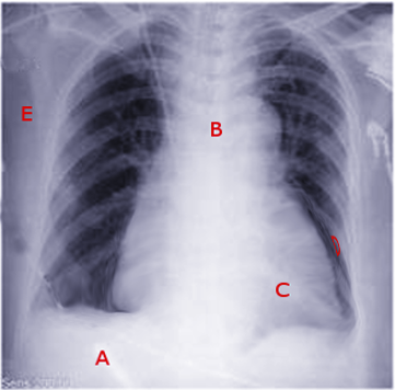
Mahabadi N, Goizueta AA, Bordoni B. Anatomy, Thorax, Lung Pleura And Mediastinum. StatPearls. 2023 Jan:():     [PubMed PMID: 30085590]


[6]

Rivard AB, Kortz MW, Burns B. Anatomy, Head and Neck: Internal Jugular Vein. StatPearls. 2024 Jan:():     [PubMed PMID: 30020630]


[7]

Hojsak I, Lacaille F, Gupte GL, Köglmeier J. Central Line in Long-term Parenteral Nutrition in Children: A European Survey. Journal of pediatric gastroenterology and nutrition. 2018 Sep:67(3):409-413. doi: 10.1097/MPG.0000000000002064. Epub     [PubMed PMID: 29916949]

Level 3 (low-level) evidence

[8]

Lambert I, Tarima S, Uhing M, Cohen SS. Risk Factors Linked to Central Catheter-Associated Thrombosis in Critically Ill Infants in the Neonatal Intensive Care Unit. American journal of perinatology. 2019 Feb:36(3):291-295. doi: 10.1055/s-0038-1667377. Epub 2018 Aug 6     [PubMed PMID: 30081400]


[9]

Paik P, Arukala SK, Sule AA. Right Site, Wrong Route - Cannulating the Left Internal Jugular Vein. Cureus. 2018 Jan 9:10(1):e2044. doi: 10.7759/cureus.2044. Epub 2018 Jan 9     [PubMed PMID: 29541565]


[10]

Soffler MI, Hayes MM, Smith CC. Central venous catheterization training: current perspectives on the role of simulation. Advances in medical education and practice. 2018:9():395-403. doi: 10.2147/AMEP.S142605. Epub 2018 May 25     [PubMed PMID: 29872360]

Level 3 (low-level) evidence

[11]

Arafa R, Khammas H, Agrawal A. Guidewire-induced asystole complicating a right internal jugular catheter placement in a patient with pre-existing left bundle branch block: A case report. The journal of vascular access. 2019 Mar:20(2):217-221. doi: 10.1177/1129729818783966. Epub 2018 Jul 9     [PubMed PMID: 29984628]

Level 3 (low-level) evidence

[12]

Amir R, Knio ZO, Mahmood F, Oren-Grinberg A, Leibowitz A, Bose R, Shaefi S, Mitchell JD, Ahmed M, Bardia A, Talmor D, Matyal R. Ultrasound as a Screening Tool for Central Venous Catheter Positioning and Exclusion of Pneumothorax. Critical care medicine. 2017 Jul:45(7):1192-1198. doi: 10.1097/CCM.0000000000002451. Epub     [PubMed PMID: 28422778]


[13]

Weekes AJ, Keller SM, Efune B, Ghali S, Runyon M. Prospective comparison of ultrasound and CXR for confirmation of central vascular catheter placement. Emergency medicine journal : EMJ. 2016 Mar:33(3):176-80. doi: 10.1136/emermed-2015-205000. Epub 2015 Oct 7     [PubMed PMID: 26446313]


[14]

Cleff C, Boensch M, Eifinger F, Hinkelbein J. [Correct positioning of central venous catheters in pediatrics : Are current formulae really useful?]. Der Anaesthesist. 2018 Jul:67(7):519-524. doi: 10.1007/s00101-018-0446-1. Epub     [PubMed PMID: 29736556]