Introduction
The upper limb comprises many muscles which are organized into anatomical compartments. These muscles act on the various joints of the hand, arm, and shoulder, maintaining tone, providing stability and allowing precise fluid movement.
Axioappendicular groups of muscles arise from the axial skeleton to act upon the pectoral girdle. Scapulohumeral muscles originate from the scapula and insert into the proximal humerus. Included in this category are the rotator cuff muscles which provide stability to the glenohumeral joint. In the arm, the muscles of the anterior compartment are involved in flexion of the forearm, and the posterior comprises of the forearm extensors. Similarly, the anterior compartment of the forearm contains the flexors of the hand and posterior has extensors. The hand is divided into the thenar, the hypothenar, the adductor compartment, as well as the short muscles of the hand.[1][2][3][4]
Structure and Function
Shoulder and Upper limb muscles are specialized to perform functions of pressure and manipulation of objects. Skeletal muscle is formed by myofibers which contain millions of myofibrils, and each of them is formed by sarcomeres. Sarcomeres are the contractile unit, therefore skeletal muscle fibers are completely dedicated to generating force. Skeletal muscle also carries out multiple functions such as voluntary locomotion, the protection of internal organs, generation of heat, and assisting in postural behavior. [5][6]
The muscle mass relies on the balance between synthesis and degradation of proteins, the regulation of this process is sensitive to several factors such as nutritional status, hormone levels, physical activity, underlying diseases, injuries, among others.[7]
Embryology
Mesenchyme (mesodermal in origin) condenses into sets of dermatomes and myotome complexes. Myotomes migrate into the developing limb buds, to give rise to myoblasts. Elongation of the limb buds, along with muscle formation from myoblasts, compartmentalizes the muscles into their respective muscle groups.
Blood Supply and Lymphatics
The arterial supply to the muscles of the upper limb is primarily from the axillary artery (of subclavian artery) and its branches. The brachial artery supplies to the anterior compartment of the arm, and the profunda brachii supplies the posterior. In the arm, the radial artery supplies the lateral forearm and the ulnar is responsible for the medial aspect. Beyond the wrist, the radial and ulnar arteries form the superficial and deep palmar arterial arches. The deoxygenated blood drains into the cephalic vein and the basilic vein. Lymphatics of the right upper limb drain into the right lymphatic duct, and the left drains into the thoracic duct.[4][8][9]
Nerves
The innervation of the muscles of the upper limb is through branches of the brachial plexus which is composed of the ventral rami of C5 through T1 nerve roots.[10][11]
Muscles
Anterior Axioappendicular Muscles (Thoracoappendicular Muscles)[12]
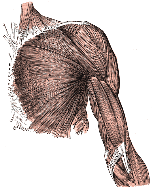
Pectoralis major
- Function: flexion, adduction, medial rotation of the humerus.
- Origin: clavicular head: medial clavicle anteriorly, sternocostal head: anterior sternum and costal cartilages of ribs 1 to 6 as well as external oblique aponeurosis
- Insertion: lateral edge of intertrabecular groove of humerus
- Innervation: medial pectoral nerve (C8, T1) lateral pectoral nerve (C5, C6, C7) of brachial plexus
Pectoralis minor
- Function: Depression of shoulder, protraction of scapula
- Origin: Third, fourth, fifth ribs close to their respective costal cartilages
- Insertion: Coracoid process
- Innervation: Medial pectoral nerve (C8, T1)
Subclavius
- Function: Depression and stabilization of clavicle
- Origin: First rib medially
- Insertion: Middle of clavicle, inferiorly
- Innervation: Nerve to subclavius (C5, C6)
Serratus anterior
- Function: Protraction of scapula, rotation of scapula
- Origin: Lateral first to the eighth rib
- Insertion: anterior scapula, medially
- Innervation: long thoracic nerve (C5, C6, C7)
Posterior Axioappendicular Muscles
Superficial layer
Latissimus dorsi
- Function: Adduction, medial rotation, extension of humerus
- Origin: Spinous processes of seventh to 12th thoracic vertebrae, iliac crest, thoracolumbar fascia, and inferior third and fourth rib
- Insertion: Intertubercular groove of humerus
- Innervation: Thoracodorsal nerve (C5,C6,C7)
Trapezius
- Function: Elevation, depression, and retraction of the scapula, rotation of glenoid cavity
- Origin: Superior nuchal line, nuchal ligament, occipital protuberance, spinous processes of C7- T12
- Insertion: Spine of scapula, acromion, and lateral clavicle
- Innervation: CN XI
Deep Layer
Levator scapulae
- Function: Adduction, medial rotation, extension of humerus
- Origin: Transverse processes of C1 through C4 vertebrae
- Insertion: Scapula at its medial border
- Innervation: Thoracodorsal nerve (C5, C6, C7)
Rhomboid major
- Function: Retraction of scapula and depression of glenoid cavity
- Origin: Spinous processes of T2 through T5 vertebrae
- Insertion: Inferior aspect of medial scapula
- Innervation: Dorsal scapular nerve (C4, C5)
Rhomboid minor
- Function: Retraction of scapula and depression of glenoid cavity
- Origin: Nuchal ligament as well as spines of C7 and T1 vertebrae
- Insertion: Superior aspect of medial scapula
- Innervation: Dorsal scapular nerve (C4, C5)
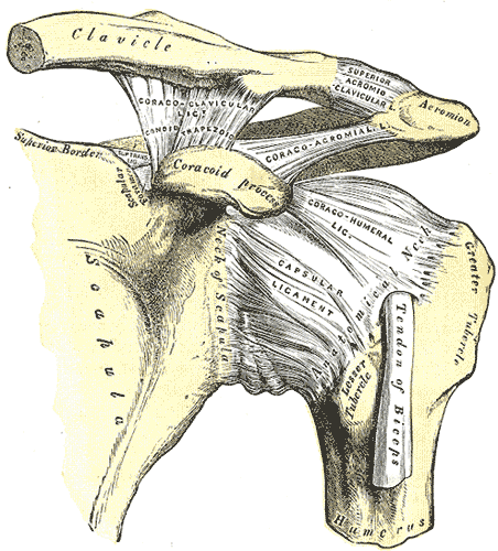
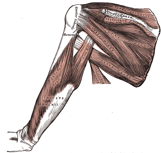
Scapulohumeral (Intrinsic Shoulder Muscles)
Deltoid
- Function: Anterior part: flexion and medial rotation of arm, middle part: abduction of arm, posterior part: extension and lateral rotation of arm
- Origin: Lateral clavicle, acromion and scapular spine
- Insertion: Deltoid tuberosity
- Innervation: Axillary nerve (C5, C6)
Teres major
- Function: Adduction and medial rotation of arm
- Origin: Posterior surface of scapula at its inferior angle
- Insertion: Intertubercular groove on its medial aspect
- Innervation: Lower scapular nerve (C5, C6)
Supraspinatus
- Function: Initiation of arm abduction
- Origin: Posterior scapula, superior to the scapular spine
- Insertion: Superior aspect of the greater tubercle
- Innervation: Suprascapular nerve (C5, C6)
- Part of rotator cuff muscles
Infraspinatus
- Function: Lateral rotation of arm
- Origin: Posterior scapula, inferior to the scapular spine
- Insertion: Greater tubercle of humerus, between supraspinatus and teres minor insertion
- Innervation: Suprascapular nerve (C5, C6)
- Part of rotator cuff muscles
Teres minor
- Function: Lateral rotation of arm
- Origin: Posterior surface of scapula at its inferior angle
- Insertion: Inferior aspect of the greater tubercle
- Innervation: Axillary nerve (C5, C6)
- Part of rotator cuff muscles
Subscapularis
- Function: Adduction and medial rotation of the arm
- Origin: Anterior aspect of scapula
- Insertion: Lesser tubercle of humerus
- Innervation: Subscapular nerves (C5, C6, C7)
- Part of rotator cuff muscles
*Rotator cuff muscles: supraspinatus, infraspinatus, teres minor, subscapularis
Muscles of Anterior Compartment of Arm (Flexors of Arm)
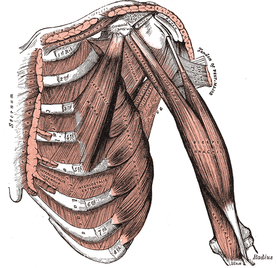
Biceps brachii
- Function: Major flexion of forearm, supination of forearm, resists dislocation of shoulder
- Origin: Short head originates from the coracoid process. The long head is from the supraglenoid tubercle of scapula
- Insertion: Radial tuberosity and forearm fascia (as bicipital aponeurosis)
- Innervation: Musculocutaneous nerve (C5, C6)
Brachialis
- Function: Flexion of forearm
- Origin: Distal anterior humerus
- Insertion: Coronoid process and ulnar tuberosity
- Innervation: musculocutaneous nerve (C5, C6, C7 small contribution)
Coracobrachialis
- Function: Flexion and adduction of arm
- Origin: Coracoid process
- Insertion: Middle of the humerus, on its medial aspect
- Innervation: Musculocutaneous nerve (C5, C6, C7)
Muscles of Posterior Compartment of Arm (Extensors of Arm)
Triceps brachii
- Function: Major extensor of forearm, resists dislocation of shoulder
- Origin: Lateral head: above the radial groove, medial head: below the radial groove, long head: infraglenoid tubercle of scapula
- Insertion: Olecranon process of ulna and forearm fascia
- Innervation: Radial nerve (C6,C7,C8)
Anconeus
- Function: Extension of forearm, stabilization of elbow joint
- Origin: Lateral epicondyle of humerus
- Insertion: Olecranon process and posterior ulna
- Innervation: Radial nerve (C7, C8, T1)
Muscles of Anterior Compartment of Forearm (Flexors of Forearm)
Superficial layer
Pronator teres
- Function: Pronation of radio-ulnar joint
- Origin: Coronoid process and medial epicondyle of humerus
- Insertion: Lateral surface of the radius
- Innervation: Median nerve (C6, C7)
Flexor carpi radialis
- Function: Flexion and adduction at the wrist
- Origin: Medial epicondyle of humerus
- Insertion: Base of second metacarpal
- Innervation: Median nerve (C6, C7)
Palmaris longus
- Function: Flexion at the wrist, tensing of the palmaris aponeurosis
- Origin: Medial epicondyle of humerus
- Insertion: Flexor retinaculum
- Innervation: Median nerve (C7, C8)
Flexor carpi ulnaris
- Function: Flexion and adduction at the wrist
- Origin: Medial epicondyle of humerus and olecranon
- Insertion: Pisiform, hook of hamate and fifth metacarpal
- Innervation: Median nerve (C7, C8)
Intermediate Layer
Flexor digitorum superficialis
- Function: Flexion of the proximal interphalangeal joint of the second, third, fourth, and fifth finger. Also has a weaker flexion action on the metacarpophalangeal joints of the same fingers
- Origin: Medial epicondyle, coronoid process, and anterior radius
- Insertion: Second, third, fourth, and fifth middle phalanges
- Innervation: Median nerve (C7, C8, T1)
Deep Layer
Flexor digitorum profundus
- Function: Flexion of the distal interphalangeal joint of the second, third, fourth, and fifth finger
- Origin: Medial and anterior surface of proximal ulna and interosseous membrane
- Insertion: Second, third, fourth, and fifth distal phalanges
- Innervation: Ulnar nerve (C8, T1) for the medial part, anterior interosseous nerve (C8,T1) for the lateral
Flexor pollicis longus
- Function: Flexion of the interphalangeal joint of the thumb
- Origin: Anterior aspect of radius as well as interosseous membrane
- Insertion: Base of distal phalanx of thumb
- Innervation: Anterior interosseous nerve (C7, C8)
Pronator quadratus
- Function: Pronator of forearm
- Origin: Anterior aspect of distal ulna
- Insertion: Anterior aspect of distal radius
- Innervation: Anterior interosseous nerve (C7, C8)
Brachioradialis
- Function: Weak flexor of the forearm
- Origin: Proximal supracondylar ridge on humerus
- Insertion: Lateral surface of distal end of radius
- Innervation: Radial nerve (C5, C6, C7)
Muscles of Posterior Compartment of Forearm
Superficial
Extensor carpi radialis longus
- Function: Extension and abduction of the wrist
- Origin: Proximal supracondylar ridge on humerus
- Insertion: Dorsal base of second metacarpal
- Innervation: Radial nerve (C6, C7)
Extensor carpi radialis brevis
- Function: Extension and abduction of the wrist
- Origin: Lateral epicondyle of humerus
- Insertion: Dorsal base of third metacarpal
- Innervation: Deep branch of the radial nerve (C7, C8)
Extensor digitorum
- Function: Extension of the proximal interphalangeal joint of the second, third, fourth, and fifth finger. Also has a weaker extension action on the metacarpophalangeal joints of the same fingers
- Origin: Lateral epicondyle of humerus
- Insertion: Extensor expansions on dorsal aspect of second, third, fourth, and fifth middle and distal phalanges
- Innervation: Posterior interosseous nerve (C7, C8)
Extensor digiti minimi
- Function: Extension of the little finger at metacarpophalangeal joint and interphalangeal joint
- Origin: Lateral epicondyle of humerus
- Insertion: Extensor expansion on dorsal aspect of fifth phalanx
- Innervation: Posterior interosseous nerve (C7, C8)
Extensor carpi ulnaris
- Function: Extension and adduction of the wrist
- Origin: Lateral epicondyle of humerus and posterior ulna
- Insertion: Fifth metacarpal base
- Innervation: Posterior interosseous nerve (C7, C8)
Deep Layer
Extensor indicis
- Function: Extension of the index finger
- Origin: Dorsal surface of distal ulna and interosseous membrane
- Insertion: Extensor expansion of second finger
- Innervation: Posterior interosseous nerve (C7, C8)
Supinator
- Function: Supination of the forearm
- Origin: Lateral epicondyle and supinator crest of ulna
- Insertion: Lateral surface of radius
- Innervation: Deep branch of radial nerve (C7, C8)
Abductor pollicus longus
- Function: Abduction of the thumb by acting on the carpometacarpal joint and the metacarpophalangeal joint
- Origin: Dorsal aspects of proximal radius, ulna, and interosseous membrane
- Insertion: Base of first metacarpal
- Innervation: Posterior interosseous nerve (C7, C8)
Extensor pollicus longus
- Function: Extension of the thumb by acting on the carpometacarpal joint, the metacarpophalangeal joint, and the interphalangeal joint.
- Origin: Dorsal aspects of middle ulna and interosseous membrane
- Insertion: Distal phalanx of 1st finger
- Innervation: Posterior interosseous nerve (C7, C8)
Extensor pollicus brevis
- Function: Extension of the thumb by acting on the carpometacarpal joint and the metacarpophalangeal joint
- Origin: Dorsal aspects of middle radius and interosseous membrane
- Insertion: Distal phalanx of 1st finger
- innervation: Posterior interosseous nerve (C7, C8)
Intrinsic Muscles of Hand
Thenar muscles
Opponens pollicus
- Function: Opposition of the thumb
- Origin: Flexor retinaculum and tubercle of trapezium
- Insertion: Lateral aspect of first metacarpal
- Innervation: Recurrent branch of median nerve (C8, T1)
Abductor pollicus brevis
- Function: Abduction of the thumb at the metacarpophalangeal joint
- Origin: Flexor retinaculum and tubercle of scaphoid
- Insertion: Lateral aspect of proximal phalanx of first finger
- Innervation: Recurrent branch of median nerve (C8, T1)
Flexor pollicus brevis
- Function: Flexion of the thumb at the metacarpophalangeal joint
- Origin: Flexor retinaculum and tubercle of trapezium
- Insertion: Lateral aspect of proximal phalanx of first finger
- Innervation: Recurrent branch of median nerve (C8, T1)
Adductor Compartment
Adductor pollicus
- Function: Adduction of the thumb
- Origin: Second, third metacarpal, and capitate
- Insertion: Proximal phalanx and extensor expansion of 1st finger
- Innervation: Deep branch of ulnar nerve (C8, T1)
Hypothenar Muscles
Abductor digiti minimi
- Function: Abduction of the little finger at the metacarpophalangeal joint
- Origin: Pisiform
- Insertion: Medial aspect of proximal phalanx of fifth finger
- Innervation: Deep branch of ulnar nerve (C8, T1)
Flexor digiti minimi brevis
- Function: Flexion of the little finger at the metacarpophalangeal joint
- Origin: Flexor retinaculum and hook of hamate
- Insertion: Medial aspect of proximal phalanx of fifth finger
- Innervation: Deep branch of ulnar nerve (C8, T1)
Opponens digiti minimi
- Function: Opposition of the little finger
- Origin: Flexor retinaculum and hook of hamate
- Insertion: Medial aspect of fifth metacarpal
- Innervation: Deep branch of ulnar nerve (C8, T1)
Short Muscles
Lumbricals
- Function: Flexion of the metacarpophalangeal joints with extension of the interphalangeal joints
- Origin: Arise from tendons of flexor digitorum profundus. First 2 are unipennate, and the third and fourth are bipennate
- Insertion: Extensor expansions of second, third, fourth, and fifth finger
- Innervation: Median nerve (C8, T1) for the lateral 2 lumbricals, deep branch of ulnar nerve (C8, T1) for the medial 2 lumbricals
Dorsal interossei
- Function: Abduction of the second, third, and fourth finger away from the axial line
- Origin: Adjacent metacarpals
- Insertion: Extensor expansions and proximal phalanges of the second, third, and fourth fingers
- Innervation: Deep branch of ulnar nerve (C8, T1)
Palmar interossei
- Function: Adduction of the second, third, and fourth finger towards the axial line
- Origin: Palmar surfaces of second, fourth, and fifth metacarpals
- Insertion: Extensor expansions and proximal phalanges of the second, fourth, and fifth fingers
- Innervation: Deep branch of ulnar nerve (C8, T1)
Physiologic Variants
The biceps brachii is one of the most variable muscles in the human body, this muscle can have more than two heads arising from humerus at the insertion of the coracobrachialis or neck of the humerus. Some reports describe supernumerary bicipital heads fluctuating from 3 to 7 in different groups, being the 3 heads variant the most common type.
Palmaris longus variations have also been described. The reversed palmaris longus is a polymorphic variation where the muscle is fleshy distally and tendinous proximally. Other case reports have documented a variation in its distal attachment of the muscle to the tendon of the adductor pollicis brevis, and long flexor; or to the flexor carpis ulnaris muscle, and muscle of the hypothenar eminence; as well as to the fascias of the forearm or the hypothenar eminence, and at the metacarpophalangeal joint. [13]
Surgical Considerations
Upper extremity injuries are usually treated in non-operative modalities such as medications, splints, injection, physical therapy. The surgical consideration of the muscles of the upper limb depends on the underlying condition.
The surgical purpose in upper limb dysfunction due to brain injury is to decrease muscle spasticity, correct joint contractures, as well as enhance the appearance, and function of the extremity. Some of the approaches to increase or decrease the muscle tone imply the peripheral nerve surgery or lengthening hyperactive muscles.
In patients with spinal cord injuries some of the surgical techniques are aimed to restore elbow rectification, and grasp of the upper limb.
Some injuries where fractures and destruction of soft tissues (skin and muscles) are present, a multidisciplinary approach is required in which the orthopedic surgeons work with plastic surgeons to treat open fractures of the extremities, osteomyelitis, or unstable scars correctly.
Clinical Significance
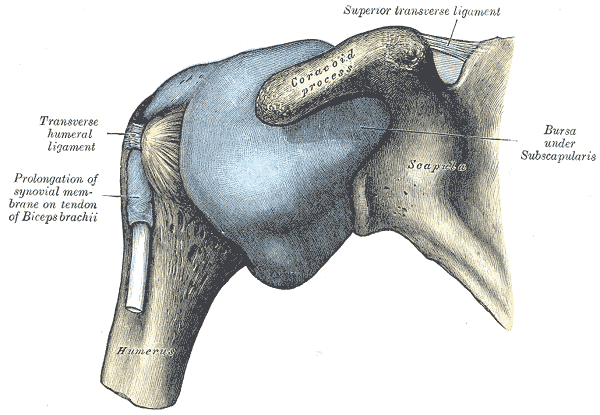
Rotator Cuff Disease
Involves impingement, tendonitis, as well as tearing of the tendons of the muscles of the rotator cuff. Majority of the cases involve the tendon of the supraspinatus muscle. This is thought to be due to its poor blood supply. The patient complains of pain, especially while lying down on the affected arm or when doing overarm activities. In the clinic, it can be tested by the Hawking’s test and Neer test. The drop test is confirmatory. An MRI is also advised to rule out or confirm a tendon tear. Treatment depends on severity. Management involves NSAIDs, physiotherapy, and arthroscopic repair.
Poland Syndrome[14]
This is the congenital ipsilateral absence or hypoplasia of pectoralis major and pectoralis minor muscles with hypoplasia of the corresponding ribs. It is hypothesized to be caused by an in-utero defect of blood supply to the developing chest. Poland syndrome is commonly associated with defects in breast and/or upper limb development.
Winging of the Scapula
Denervation of the serratus anterior muscle causes palsy of the long thoracic nerve. This causes lateral and posterior movement of the scapula, away from the underlying ribs, giving it a wing-like appearance.
Epicondylitis
Lateral epicondylitis (tennis elbow) is caused by a combination of repetitive or sustained contraction of the extensor muscles of the forearm leading to inflammation of the common extensor origin. Medial epicondylitis (golfers elbow) is due to repetitive or sustained contraction of the flexor muscles of the forearm leading to inflammation of the common flexor origin. Patients present with pain and tenderness over the affected epicondyle that worsens with extension (in the case of lateral epicondylitis) or flexion (when suspecting medial epicondylitis). Treatment involves avoiding exacerbating activities, physical therapy, and pain relief.
Other Issues
Traumatic injury, malignancy, infection, or congenital deformity of the upper extremity can lead to amputation. Surgeons have different surgical options to improve the potential of using prosthetic technologies for this group of patients. Targeted muscle reinnervation is a surgical procedure to enhance the control of myoelectric upper limb prostheses, and it helps to prevent and treat painful postamputation neuromas. This technique was originally described for transhumeral amputations and shoulder disarticulations but nowadays it has also been applied in the treatment of transtibial, transfemoral, transradial, and partial hand amputees. [15][16]