Continuing Education Activity
Total knee arthroplasty (TKA) is one of the most cost-effective and consistently successful surgeries in orthopedics. It provides reliable outcomes for patients suffering from end-stage degenerative knee osteoarthritis. Specifically, it can alleviate pain, restore function, and lead to an improved quality of life. This activity reviews the indications, contraindications, and techniques involved in performing total knee arthroplasty and highlights the role of the interprofessional team in the care of patients undergoing this procedure.
Objectives:
- List the indications for total knee arthroplasty.
- Describe the technique involved in performing total knee arthroplasty.
- List the complications associated with total knee arthroplasty.
- Explore interprofessional team strategies for improving care coordination and communication to advance the management of patients requiring total knee arthroplasty.
Introduction
Total knee arthroplasty (TKA) is one of the most cost-effective and consistently successful surgeries performed in orthopedics. Patient-reported outcomes are shown to improve dramatically with respect to pain relief, functional restoration, and improved quality of life. TKA provides reliable outcomes for patients suffering from end-stage, tri-compartmental, degenerative osteoarthritis (OA). While OA affects millions of Americans, the knee is the most commonly affected joint plagued by this progressive condition which is hallmarked by a gradual degeneration and loss of articular cartilage.
Estimates project the annual incidence of symptomatic knee OA at 240 per 100,000 patients per year, and about 400,000 primary TKA surgeries are performed annually in the United States. The most common clinical diagnosis associated with TKA is primary OA, but other potential underlying diagnoses include inflammatory arthritis, fracture (post-traumatic OA and/or deformity), dysplasia, and malignancy.[1][2][3]
Anatomy and Physiology
The knee is comprised of 2 separate joints: the tibiofemoral and patellofemoral joints.[4][5][6]
Patellofemoral Joint
The patellofemoral joint (PFJ) functions to increase the lever arm of the extensor mechanism. The patella transmits the tensile forces generated by the quadriceps tendon to the patellar tendon. The maximum contact force between the patella and femoral trochlea occurs at 45 degrees of knee flexion, and joint reaction forces reach 7-times body weight in the position of deep squatting.
The quadriceps muscles provide dynamic stability of the PFJ, and passive anatomic restraints include the following:
- Medial patellofemoral ligament: Primary passive restraint against lateral translation at 20 degrees of flexion
- Medial patellomeniscal ligament: Contributes 10% to 15% of the total restraining force
- Lateral retinaculum: Provides 10% of the total restraining force
Tibiofemoral Articulation
The tibiofemoral articulation transmits body weight from the femur to the tibia and generates joint reaction forces of 3 and 4-times body weight during walking and climbing, respectively. Motion occurs in the sagittal plane from 10 degrees of hyperextension to about 140 to 150 degrees of hyperflexion. Extremes of flexion are often limited secondary to direct contact between the posterior thigh and calf. The tibiofemoral contact point and femoral center of rotation move posteriorly with increasing degrees of flexion in order to optimize knee flexion prior to impingement. Normal gait only requires a range of motion (ROM) from 0 to 75 degrees.
Knee stability in the coronal plane is provided by the lateral collateral ligament (LCL), which resists varus stresses, and the medial collateral ligament, which resists valgus stress forces. In addition, the anterior cruciate ligament (ACL) and posterior cruciate ligament (PCL) provide resistance to anteriorly directed and posteriorly directed forces at the knee, respectively. Resistance to external rotatory forces is provided by the posterolateral corner structures (PLC).
Indications
Once considered a procedure reserved for the elderly, low-demand patient population, primary TKA is offered more frequently and provides consistent positive outcomes in younger cohorts of patients. In general, the most common underlying diagnosis associated with performing TKAs across all patient age groups is primary, end-stage, tri-compartmental osteoarthritis.[7][8][9]
TKA is an elective procedure that is, in most cases, reserved for patients experiencing chronic, debilitating symptoms that continue to persist despite exhaustion of all conservative and nonoperative treatment modalities.
Contraindications
TKA is contraindicated in the following clinical scenarios:
- Local knee infection or sepsis
- Remote (extra-articular), active, ongoing infection or bacteremia
- Severe cases of vascular dysfunction
Equipment
TKA prosthesis designs have been evolving since the 1950s, beginning with Walldius’ design of the first hinged-knee replacement. In the early 1970s, the total condylar prosthesis (TCP) was the first TKA prosthesis designed to resurface all 3 compartments of the knee. The TCP was a posterior-stabilized design. The 4 main categories of TKA prosthesis designs are listed below in the order of increasing levels of constraint by design.[10][11][12]
Cruciate-Retaining
The cruciate-retaining TKA prosthesis depends on an intact PCL to provide stability in flexion. Thus, its use is contraindicated in patients with pre-existing or intra-operatively recognized PCL insufficiency. Caution is given to any patient presenting with at least moderate instability in any plane of motion, especially PLC instability patients. PLC instability predisposes the native PCL in a cruciate-retaining TKA to abnormally high stresses and forces, ultimately leading to early failure and TKA instability requiring revision. Cruciate-retaining TKA is contraindicated in patients suffering from inflammatory arthritic conditions, given the increased risk of early PCL attenuation (e.g., rheumatoid arthritis).
Proposed advantages of the cruciate-retaining TKA design include:
- Avoidance of tibial post-cam impingement and dislocation
- Retaining more normal anatomy theoretically resembles normal knee kinematics
- Preserved bone stock (less distal femur resected compared to PS TKA prosthesis)
- Native PCL proprioception
Proposed disadvantages of the cruciate-retaining TKA design include:
- A tight PCL can lead to early/accelerated polyethylene wear
- Loose/ruptured PCL results in flexion instability and possible subluxation/dislocation
Multiple meta-analyses have demonstrated satisfactory survivorship and similar outcomes comparing the cruciate-retaining and posterior-stabilized TKA prosthesis designs.
Posterior-Stabilized
The posterior-stabilized TKA design is slightly more constrained and requires the surgeon to sacrifice the PCL. The femoral component contains a cam that is designed to engage the tibial polyethylene post as the knee flexes.
Proposed advantages of the posterior-stabilized TKA design include:
- Facilitates overall balancing of the knee in the setting of an absent PCL
- Theoretically, better knee flexion
- Lower ranges of axial rotation and condylar translation
Proposed disadvantages of the posterior-stabilized TKA design include:
- Cam jump that can result secondary to a loose flexion gap or in knee hyperextension
- Patellar clunk syndrome
- Tibial post wear and/or fracture
Constrained Nonhinged Design
The constrained nonhinged prosthesis employs a larger tibial post and deeper femoral box, yielding more stability and constraint (within 2 to 3 degrees) in both varus-valgus and internal-external rotatory planes. Indications include collateral ligament attenuation or deficiency, flexion gap laxity, and moderate bone loss in the setting of neuropathic arthropathy. Downsides to this design include not only an increased risk of earlier aseptic loosening secondary to the increased inter-component constraint but also the requirement of more femoral bone resection to accommodate the components.
Constrained Hinged Design
The constrained hinged design is comprised of linked femoral and tibial components. Rotating hinge options allow the tibial bearing to rotate around a yoke that theoretically mitigates the risk of aseptic loosening at the expense of increasing levels of prosthetic constraint. Indications include global ligamentous deficiencies, resections in the setting of tumors, and massive bone loss in the setting of a neuropathic joint.
Other Component Considerations
Modularity and mobile bearing designs are other noteworthy additional prosthetic design considerations.
Mobile bearing designs allow polyethylene rotation on the tibial baseplate. Although this design concept remains controversial in terms of its generation of reproducibly superior patient-reported outcome measures, advocates cite its utilization and relative indications in younger patient populations secondary to improved wear rates. However, one notable disadvantage includes the potential for bearing spin-out, which is seen especially in the setting of a loose flexion gap.
All-polyethylene tibial base plates contrast the conventional metal tray with polyethylene inserts (i.e., tibial component modularity) that allow surgeons more flexibility for intra-operative adjustments for fine-tuning TKA stability. A surgeon is able to upsize or downsize the polyethylene after the final tibial implant fixation has been achieved between the metal implant and cement (or bone) interfaces. This allows for a final check and balance step, which many TKA surgeons appreciate. In contrast, advocates for the all-polyethylene base plates cite significant cost savings and decreased rates of osteolysis when comparing TKA cohorts, especially in the elderly TKA patient populations.[13][14]
Preparation
Nonoperative Treatment Modalities
According to the 2011 American Academy of Orthopaedic Surgeons (AAOS), Evidence-Based Clinical Guidelines for the treatment of symptomatic hip or knee osteoarthritis, strong or moderately strong recommendations for nonoperative treatment modalities include weight loss, physical activity, physical therapy programs, and NSAIDs and/or tramadol. Other modalities that were not supported by moderate or strong evidence but are often considered reasonable alternative treatment options include but are not limited to acupuncture, chondroitin supplementation, hyaluronic acid injections, corticosteroid injections, lateral wedge insoles, and offloading braces.[15]
Preoperative Evaluation: Clinical Examination
A thorough history and physical examination are required before performing a TKA on any patient. Patients should be asked about any and all previous interventions and treatments. Prior joint replacements, arthroscopic procedures, or other surgeries around the knee should be considered. Old surgical scars can affect the planned surgical approach. In addition, patients with a history of prior injuries or procedures can present with mechanical axis deformities, retained hardware, or knee instability in any plane. A multitude of factors can impact the TKA prosthesis of choice that is most appropriate for the patient.
We recommend each patient pursuing elective TKA surgery first receive a comprehensive medical evaluation with any appropriate medical optimization tests performed before the TKA procedure. A surgeon must consider the relevant risks and potential benefits of performing TKA on a case-by-case basis.
Physical examination includes evaluation of the overall mechanical axis of the limb. It is critical to ensure hip pathology is either ruled out or at least considered before performing any surgery around the knee. The vascular status of the limb should also be assessed by observing the skin for any chronic venous stasis changes, cellulitis, or even wounds/ulcerations that may be present on the extremity. Distally, the pulses should be symmetric and palpable. Consideration should be given to a vascular surgeon consultation in the preoperative setting in any patient presenting with peripheral vascular disease (PVD). The surgeon should also be aware of the possibility of PVD presenting as knee pain out of proportion in the setting of relatively benign radiographs.
Preoperative range of motion should be noted at the knee and adjacent joints (hip, ankle). The soft tissues should be examined for evidence of gross atrophy, overall symmetry, and ligamentous stability in all planes at the knee joint. It is essential to document the presence of any laxity in the varus/valgus plane and the ability to correct the deformity. These parameters help prepare the surgeon for soft tissue releases that may be required to facilitate mechanical axis correction, as well as plan for additional bone resection that may be needed in the setting of significant contractures.
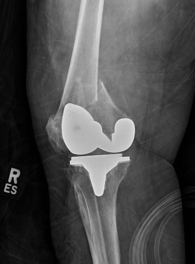
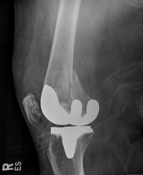
Preoperative Evaluation: Radiographs
Preoperative radiographs, including a weight-bearing anteroposterior (AP) view, are evaluated for overall mechanical alignment, the presence of deformity, and bone loss. The tibiofemoral angle can help estimate the magnitude of coronal deformity. The femoral resection angle is calculated as the difference between the mechanical and anatomic axis of the femur. The lateral view of the knee is essential for appreciating the native posterior slope of the proximal tibia as well as the presence of posterior osteophytes on the femoral condyles.
The patellofemoral radiographic view is not necessary for TKA templating but allows the surgeon to evaluate the magnitude of patellofemoral arthritis and deformity. In cases of advanced patellofemoral deformity, osteophyte removal may be needed prior to attempting to evert the patella during the procedure. In addition, a surgeon can plan for a possible lateral release to improve patellar tracking.
Technique or Treatment
Surgical Approaches
The most common approaches for the standard primary TKA procedure include the medial parapatellar, midvastus, and subvastus approaches. The medial parapatellar approach is commonly utilized and entails proximal dissection through a medial cuff of the quadriceps tendon to facilitate superior tissue quality closure at the conclusion of the procedure. Distally, a meticulous, continuous medial subperiosteal dissection sleeve is performed while maintaining intimacy with the proximal tibial bone. The extent of dissection is often dictated by the anticipated amount of deformity to be corrected. In general, this medial release is aggressive in cases of severe varus deformity and most minimal in cases of moderate to advanced valgus knee deformity. The medial meniscus is also resected with this sleeve of soft tissue.[16][17][18]
Alternatives to the standard medial parapatellar arthrotomy include the midvastus and subvastus approaches. The midvastus approach spares the quadriceps tendon. Instead, the vastus medialis obliquus (VMO) muscle belly is dissected along a trajectory directed toward the superomedial aspect of the proximal pole of the patella.
The subvastus approach also spares the quadriceps tendon and lifts the muscle belly of the VMO off the intermuscular septum. The subvastus approach preserves the vascularity of the patella and is cautioned as it can limit exposure in particularly challenging cases or in particularly obese patients.
Procedural Steps
Depending on surgeon preference, the specific order of bone resections and soft tissue releases will vary. However, a general overview of a preferred method is the goal of this technical summary of the TKA procedure.
Once the arthrotomy is complete, the patella is everted, and the knee is flexed with additional soft tissue releases required prior to achieving knee dislocation. If the surgeon elects to proceed with the femur first, an intramedullary (IM) drill is utilized in order to gain access to the femoral canal for the utilization of a distal femoral IM jig. The angle set on the guide is based on the patient-specific preoperative evaluation (AP X-ray), generally yielding 5 or 7 degrees of valgus. Although system-specific, most surgeons prefer resecting 9 to 10 mm of the distal femur.
Next, the proximal tibia is cut utilizing an IM or extramedullary (EM) guide with the goal of cutting the bone perpendicular (or within 2 to 3 degrees of varus for surgeons aiming for an "anatomic" TKA procedure) to the tibial axis. We prefer an IM guide and a perpendicular tibial cut. The rotation is set referencing the medial one-third of the tibial tubercle (proximally) and a point slightly medial to the center of the ankle joint (distally). This alignment is also referenced with the second ray of the foot and the tibial crest.
Once the tibial cut is performed, the extension gap can be assessed. A spacer block is then inserted with the knee in full extension, and the overall balance of the knee is assessed using an alignment rod to facilitate and verify overall varus-valgus and tibial slope parameters achieved.
Next, the flexion gap is attained after utilizing an AP sizing guide that is positioned with respect to the bony landmarks on the femur (usually Whiteside’s line or the native transepicondylar axis [TEA]). Depending on the anterior or posterior referencing style of the operating surgeon, the flexion gap is set and adjusted as needed utilizing the system-specific incremental sizing adjustments available with respect to the cutting guides. Prior to making the bony cuts, the flexion gap should be visualized, and soft tissue balancing appreciated. A spacer block can facilitate this assessment. The surgeon should ensure a rectangular flexion gap will be the ultimate result after the bone resections. After satisfactory check and balancing steps are verified, the anterior, posterior, anterior chamfer and posterior chamfer cuts are made. Care is taken to protect the collateral soft tissue structures (LCL, MCL) with retractors.
Next, the intercondylar notch cut is made perpendicular to the TEA. The attention is again turned back to the proximal tibia to finish preparation, sizing, and rotational alignment. One must be cautious to avoid internal rotation and/or component overhang, which can lead to inferior TKA results. The femoral and tibial trial implants are impacted, and a provisional spacer trial is inserted. The knee is reduced and assessed for stability from 0 degrees of extension through mid-flexion stability.
If planning to resurface the patella, the resection is recommended after first appreciating the native anatomy and size of the entire patellofemoral joint. Inferior TKA outcomes can result from either over-resection, which can compromise implant bone stock and lead to patella fracture, or under-resection, which can lead to chronic postoperative pain secondary to an overstuffed PFJ.
Finally, the stability parameters are again verified, and patellar tracking is appreciated and must pass intraoperative tracking tests. Most surgeons either use a natural range of motion tracking test to ensure the TKA passes the “no thumb” test, or a towel clip technique can be used.
Patellar maltracking, most commonly occurring laterally, can most often be corrected with a standard lateral release. In more severe cases or in scenarios consistent with component malalignment, consideration should be given to the correction of component position(s).
Wound Closure
The most recent literature remains controversial with respect to the ideal position of the knee and suture material utilized during the TKA closure. Attention to detail is required, and a methodical closure is unanimously advocated. A preferred method includes closure with uni- or bi-directional barbed sutures for the arthrotomy, deep fascial, and deep dermal/subcutaneous layers. Staples or monofilament absorbable sutures can be used for the skin. A sterile dressing is then applied and left in place without being changed for the first 7 days. In addition, a minimal cotton/ace soft wrap dressing is applied to the knee for, at most, 24 hours to facilitate the appropriate balance between wound healing and postoperative movement of the knee.
Other Considerations
Topical tranexamic acid (TXA) is the preferred application while waiting for the cement to fully harden and prior to dropping the tourniquet. In addition, other controversial technical modalities in TKA include the use of a tourniquet, cementing the patella, femoral, and/or tibial components, as well as incorporating a betadine soak to the wound as part of the copious saline irrigation that is applied prior to closure of the arthrotomy and surgical wound. Preferred techniques include the use of a tourniquet, cementing all components, and saline-only copious pulsatile irrigation prior to arthrotomy closure.
Complications
TKA complications result in inferior outcomes and patient-reported satisfaction scores. Although TKA remains a reliable and reproducibly successful surgery in patients suffering from debilitating advanced degenerative arthritic knees, reports still cite that up to 1 in 5 patients who have undergone primary TKA remain dissatisfied with the outcome.[19][20][21]
Periprosthetic Fracture
TKA periprosthetic fractures (PPFs) are further characterized by the location and residual stability of the implants. Distal femur PPFs occur at a 1% to 2% rate, and risk factors include compromised patient bone quality increased constrained TKA components; and while controversial, anterior femoral notching is a potential risk factor for postoperative fracture.
Tibial PPFs occur at a 0.5% to 1% rate, and risk factors include a prior tibial tubercle osteotomy, component malposition and/or loosening, as well as utilization of long-stemmed components. Patellar PPFs occur less frequently in unresurfaced TKA cases, and incidence rates range from 0.2% up rates as high as 15% or 20%. Risk factors for fracture include osteonecrosis, technical errors in asymmetric or over-resection, and implant-related associations, including the following:
- Central, single peg implants
- Uncemented fixation
- Metal-backed components
Aseptic Loosening
TKA aseptic loosening occurs secondary to a macrophage-induced inflammatory response resulting in eventual bone loss and TKA component loosening. Patients often present with pain that is increased during weight-bearing activity and/or recurrent effusions. Patients may have minimal pain at rest or with range of motion. Serial imaging and infectious labs are required to appropriately work up these conditions, which eventually are treated with revision surgery if symptoms persist and the patient is considered a reasonable surgical candidate. The steps in aseptic loosening include particulate debris formation, macrophage-induced osteolysis, micromotion of the components, and dissemination of particulate debris.
Wound Complications
The TKA postoperative wound complication spectrum ranges from superficial surgical infections (SSIs) such as cellulitis, superficial dehiscence, and/or delayed wound healing to deep infections resulting in full-thickness necrosis resulting in returns to the operating room for irrigation, debridement (incision and drainage), and rotational flap coverage.
Periprosthetic Joint Infection
The incidence of prosthetic total knee infection (TKA PJI) following primary TKA is approximately 1% to 2%, as reported in the literature. Risk factors include patient-specific lifestyle factors (morbid obesity, smoking, intravenous [IV] drug use and abuse, alcohol abuse, and poor oral hygiene) and patients with a past medical history consisting of uncontrolled diabetes, chronic renal and/or liver disease, malnutrition, and HIV (CD4 counts less than 400). PJI is the most common reason for revision surgery.
The most common offending bacterial organisms in the acute setting include Staphylococcus aureus, Staphylococcus epidermidis, and in chronic TKA PJI cases, coagulase-negative staphylococcus bacteria. Treatment in the acute setting (less than 3 weeks after index surgery) can be limited to incision and drainage, polyethylene exchange, and retention of components. In addition, IV antibiotics are utilized for up to 4 to 6 weeks duration. Outcomes vary and are often influenced by multiple intraoperative, patient-related factors and offending bacterial organisms, but studies site a 55% successful outcome rate.
More aggressive treatments, especially in the setting of presentation beyond the acute (3 to 4-week time point), include a 1 or 2-stage revision TKA procedure with interval antibiotic spacer placement. The surgeon must ensure and document evidence of infection eradication.
Other Complications and Considerations
Other potential complications after TKA are beyond the scope of this review but include:
- TKA instability: can occur in the coronal or sagittal plane(s). Also, consideration is given for patellar maltracking or other PFJ issues (for example, overstuffing the joint) in the postoperative setting when patients complain of persistent anterior knee pain
- Extensor mechanism disruption or rupture
- Patellar clunk syndrome.
- Often occurs 12 months after TKA and is associated with popping and catching during knee extension. It is caused by nodule formation on the posterior quad tendon near its insertion on the patella. Patellar clunk syndrome is associated with posterior stabilized knee design. The cause of scar tissue formation is unknown, but the pain results from tissue entrapment in the intercondylar notch. Treatment is surgical, either arthroscopic or open debridement/synovectomy. Conservative measures are often unsuccessful. Physical therapy may help with quad strengthening after surgery but is not curative. Recurrence after surgical treatment is rare. More aggressive intervention, such as revision TKA, is often not warranted in the absence of component malposition.
- Stiffness
- Vascular injury and bleeding
- Peroneal nerve palsy
- One of the most common complications after TKA is to correct valgus deformity. During soft tissue balancing of a valgus knee, the iliotibial band preferentially affects the extension space more than the flexion space and inserts on Gerdy's tubercle. The popliteus preferentially affects flexion space more than extension space.
- Metal hypersensitivity
- Heterotopic ossification
Clinical Significance
TKA is one of the most successful and cost-effective procedures in all orthopedics. The procedure is most commonly performed for patients suffering from debilitating, end-stage arthritic conditions of the knee. TKA is one of the most successful and cost-effective procedures in all orthopedics. The procedure is most commonly performed for patients suffering from debilitating, end-stage arthritic conditions of the knee. Once considered a procedure limited to the elderly, low-demand patients, TKA is becoming an increasingly popular procedure performed in younger patient populations. Between 1991 and 2010, the annual primary TKA volume in the US Medicare population alone increased by 161.5%, from more than 93,000 to more than 226,000 cases.
Enhancing Healthcare Team Outcomes
After total knee replacement, an interprofessional team is necessary to prevent and manage the comorbidities. Many of the patients are frail and/or have cognitive impairment. Hence a dietary consult is necessary. A pain consult is necessary prior to undertaking rehabilitation. However, the type of analgesic selected must allow for pain control and yet, at the same time, prevent sedation. Further, long-term pain control is not recommended as it may mask any fever infection or evidence of vascular compromise. Constipation should be managed as it is very common in this population. The pharmacist should assist the clinician in appropriate pain medication selection and management. The nurse should ensure that the patient has prophylaxis for deep vein thrombosis. Finally, the patient should be educated by the orthopedic nurse not to cross the legs, turn the leg inward or bend over to reach objects. The nurse should also assist the clinician in providing close follow up and reporting concerns about complications. The therapist should educate the patient on how to use an assistive device if the gait is unbalanced. Follow up of these patients is required until the patient is independently ambulating. [22][23][24][Level 5]
Outcomes
Although the most recent reports still cite that up to 1 in 5 patients remain dissatisfied following TKA, the risks of patient-reported dissatisfaction can be mitigated by selectively operating on patients that have had other potentially relevant clinical pathologies like hip and/or back conditions ruled out as confounding pain generators. Success following TKA results in significant improvements in patient-reported pain and functional outcome scores in the short- and long-term postoperative periods. Although the overall longevity of the TKA prosthesis is influenced by a multitude of patient-related and prosthetic technicality factors, in general, the lifespan is expected for about 15 to 20 years. Clinicians are encouraged to ensure that surgical candidates have first exhausted all nonoperative treatment modalities, as mentioned earlier in this review. As the rates of surgical procedures in the young and elderly populations continue to increase, orthopedic surgeons can expect excellent outcomes in the appropriately indicated patient populations.[25][26]