Anatomy, Thorax, Xiphoid Process


Introduction

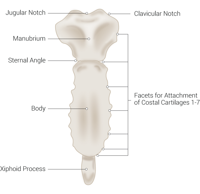
The sternum consists of 3 major parts; the manubrium, the body, and the xiphoid process, with the xiphoid process being the smallest and most distal part of the three. The manubrium is the broad, quadrangular, and most superior segment and is characterized by its superior dip known as the suprasternal notch. The body is the middle and longest part and connects to the manubrium at the sternal angle. The xiphoid process is triangular in shape and is the most distal part of the sternum.[1] See Image. Sternum Anatomy.

The term xiphoid process comes from the word "xiphos," which is of Greek origin and means straight sword describing the morphology of this bone.[2] It is approximately 2 to 5 cm in length. The xiphoid varies in shape and size; it is primarily triangular, with its base directed superiorly and the tip pointing inferiorly at the level of the T10 vertebra. The base of the xiphoid process articulates with the distal end of the sternal body forming the xiphisternal joint. Unlike the suprasternal/ jugular notch at the superior end of the manubrium of the sternum, palpating the xiphoid process can sometimes be challenging. Externally the xiphoid process is located in the epigastric region of the anterior thoracic wall.[2]

At birth, the xiphoid is purely cartilaginous and is made of 2 types of cartilage; hyaline cartilage in the proximal portion and elastic cartilage in the distal part.[3] With age, the xiphoid process ossifies; the ossification age is vastly variable.[2]

Structure and Function

The median location of the sternum in the thoracic cage and its attachment to the costal cartilage allow it to function as a protective bone to the underlying mediastinal structures.[1] The primary function of the xiphoid process is to serve as a point for muscular attachment; several muscles and ligaments attach to its different surfaces.

The anterior surface of the xiphoid process is primarily occupied by the insertion of the flat longitudinal muscles of the anterior abdominal wall. The rectus abdominis muscle serves as a strong trunk flexor and stabilizer. The aponeurosis of the internal and external oblique muscles also attaches to the anterior portion of the xiphoid process. These abdominal muscles, together with other core muscles, function to stabilize the trunk and assist in increasing intra-abdominal pressure.[4]

The posterior surface of the xiphoid process serves as an attachment point for several structures; the most important is the attachment of the diaphragmatic muscular slips providing the diaphragm with its fundamental contractile function in respiration.[2][5]

The seventh costal cartilages attach to bilateral demifacets to form the costoxiphoid/ chondroxiphoid ligament. These demifacets are located on the superior end of the xiphoid, precisely at the point where it articulates with the sternal body to form the xiphisternal joint.[6] In addition, the transverse thoracis muscle attaches to the posterior and sometimes anterior surfaces of the xiphoid process.[7] Besides these muscular attachments, the inferior surface or the tip of the xiphoid process also provides a point of attachment for the linea alba.[2]

Embryology

The embryological development of the sternum follows a highly variable pattern in terms of age and location of ossification.[1] The sternum develops from bilateral mesenchymal plates during the early weeks of gestation. These sternal plates or bands eventually fuse in the midline around the 7th week of embryonic development. This fusion starts at the cranial end of the bands and is generally complete by the 9th to 10th weeks. Until this stage, the three sternal parts would not be visible yet; as the embryonic sternum develops, the body, manubrium, and xiphoid process start to take shape.

Ossification of these three different sternal parts starts and completes at varying times following a craniocaudal direction. The manubrium and body begin to ossify around the 6th month of gestation and continue through the 1st postnatal year. Similarly, the ossification process of the xiphoid can continue up to the early years of childhood or late teenage (5th to 18th year).[8] Like the variation in shape and size of the adult form of the xiphoid process, its maturation and development vary considerably.[9][3] Interestingly, some literature marks the age of xiphoid process ossification as late as 40 to 60 years.[1]

Blood Supply and Lymphatics

The sternum is considered a highly vascular structure, receiving most of its arterial supply through perforating and sternal arterial branches located around the intercostal spaces.[10] The blood supply to the xiphoid process is derived from multiple perforating branches originating from the internal thoracic artery, also known as the internal mammary artery. The right and left internal thoracic arteries are located parallel to the sternum. Before bifurcating into the superior epigastric and musculophrenic arteries at the level of the 7th intercostal space, The internal thoracic artery gives off multiple perforating and sternal branches to supply the xiphoid process (and the sternum in general).[2] The internal thoracic artery is a branch of the first part of the subclavian artery and supplies the sternum and other mediastinal structures.[11]

The venous drainage of the xiphoid process follows a similar pattern and is collected back through the internal thoracic veins, which empty directly into the right and left brachiocephalic veins.[1]

Nerves

Due to its anatomical proximity to the intercostal spaces, the sternum, including the xiphoid process, is supplied by the intercostal nerves from the T1-T11 segments.[12] The xiphoid process is located at the level of the T6 skin dermatome of the upper thorax.[13]

Muscles

Multiple muscles are attached to different surfaces of the xiphoid process. The anterior surface of the process is primarily occupied by the insertion of the flat longitudinal muscles of the anterior abdominal wall, including the rectus abdominis muscle which serves as a strong trunk flexor and stabilizer. In addition, the aponeurosis of the internal and external oblique muscles also attaches to the anterior portion of the xiphoid process. These abdominal muscles, together with other core muscles, function to stabilize the trunk and assist in increasing intra-abdominal pressure.[4]

The posterior surface of the xiphoid process serves as an attachment point for several structures; the most important is the attachment of the diaphragmatic muscular slips providing the diaphragm with its fundamental contractile function in respiration.[5][2] In addition, the transverse thoracis muscle attaches to the posterior and sometimes anterior surfaces of the xiphoid process.[7]

Physiologic Variants

The xiphoid process is considered a variable bone, and most commonly, it varies in shape. It has been reported in the literature that the xiphoid process can take different shapes, from thin to broad. Its pointed distal end may also be curved or bifid. Some case studies have reported xiphoid foramina or a non-ossified xiphoid, which are considered unusual variations. In-depth knowledge and awareness of these variations are essential as they may lead to misdiagnosis, primarily due to their proximal anatomical relationship to vital structures in the thoracic and abdominal regions.[2]

Surgical Considerations

The xiphoid process is an important surgical landmark, especially in cardiac surgery. For example, in open-heart surgeries, this thoracic bone is used as a landmark for locating the midline while determining the exact location to place a mechanical saw before cutting through the sternum. It is imperative that the attachments of the xiphoid process are detached before inserting a saw to avoid injury to the diaphragm, lungs, or even the heart. Once the xiphoid process is detached, surgeons utilize a blunt dissection method to detach the sternal pericardial attachments by inserting a finger posterior to the sternum and bluntly dissecting from inferior to superior.

During pericardiocentesis, healthcare providers usually palpate for the xiphoid process and aim to insert the needle directly underneath the xiphoid, pointing toward the left shoulder.[14] 

Other surgical considerations of the xiphoid process include xiphoidectomy, the removal of the xiphoid process, which is commonly used to increase visualization during a total gastrectomy. This procedure has been shown to improve safety during surgery due to increased visualization of vital organs and structures.[15]

Clinical Significance

The xiphoid process is used as a landmark to determine the correct location for hand placement during cardiopulmonary resuscitation (CPR). It is essential while performing CPR not to compress at the xiphoid process as this bone is very soft, which may lead to fracture of the xiphoid and can lead to severe trauma to underlying vital organs such as the liver, heart, and diaphragm.[16][17] Knowledge of proper CPR techniques and the possibility of these fractures are imperative to all medical providers and staff.[18] 

Xiphoid syndrome is an uncommon condition presenting as painful swelling and discomfort around the xiphoid process and the epigastric region. There are limited research and case presentations related to the xiphoid syndrome. However, case studies describing this syndrome have documented that patients complain of tenderness and light pressure over the xiphoid process.[19] This uncommon syndrome is described mainly in the chiropractic literature as it is primarily considered to be associated with mechanical thoracic trauma or lifting heavy objects. It is usually described as a combination of symptoms, including diffuse epigastric pain due to inflammation of the xiphoid process. Patients also present with discomfort and pain in various body regions, the upper abdominal region, the chest, and the xiphisternal joint. The discomfort and pain can even radiate distally to the throat and the upper extremity.

The diffuse nature of pain in the xiphoid syndrome can be explained as pain referred from the inflamed joint itself, the xiphesternal joint, or radiating from the structures attached to the body of the xiphoid. Because xiphoid syndrome is an uncommon and under-described syndrome, patients can go undiagnosed for many years. Several case reports discuss the use of local anesthetic or steroidal injections around the xiphoid process leading to improvement of the symptoms of xiphodynia.[6]


(Click Image to Enlarge)
<p>Sternum Anatomy

Sternum Anatomy. The sternum anatomy includes a jugular notch, manubrium, sternal angle, body, xiphoid process, clavicular notch, and facets for attachment of costal cartilages 1-7.


Contributed by B Palmer

Details

Editor:

Bracken Burns

Updated:

3/26/2023 9:24:06 AM

References


[1]

Altalib AA, Miao KH, Menezes RG. Anatomy, Thorax, Sternum. StatPearls. 2024 Jan:():     [PubMed PMID: 31082185]


[2]

Mashriqi F, D'Antoni AV, Tubbs RS. Xiphoid Process Variations: A Review with an Extremely Unusual Case Report. Cureus. 2017 Aug 27:9(8):e1613. doi: 10.7759/cureus.1613. Epub 2017 Aug 27     [PubMed PMID: 29098125]

Level 2 (mid-level) evidence

[3]

Nam S, Cho W, Cho H, Lee J, Lee E, Son Y. Xiphoid process-derived chondrocytes: a novel cell source for elastic cartilage regeneration. Stem cells translational medicine. 2014 Nov:3(11):1381-91. doi: 10.5966/sctm.2014-0070. Epub 2014 Sep 9     [PubMed PMID: 25205841]


[4]

Adams L, Pace N, Heo A, Hunter I, Johnson AW, Mitchell UH. Internal and External Oblique Muscle Asymmetry in Sprint Hurdlers and Sprinters: A Cross-Sectional Study. Journal of sports science & medicine. 2022 Mar:21(1):120-126. doi: 10.52082/jssm.2022.120. Epub 2022 Feb 15     [PubMed PMID: 35250341]

Level 2 (mid-level) evidence

[5]

Hawkins SP, Hine AL. Diaphragmatic muscular bundles (slips): ultrasound evaluation of incidence and appearance. Clinical radiology. 1991 Sep:44(3):154-7     [PubMed PMID: 1914388]


[6]

Simpson JK, Hawken E. Xiphodynia: a diagnostic conundrum. Chiropractic & osteopathy. 2007 Sep 15:15():13     [PubMed PMID: 17868466]


[7]

Hogerzeil DP, Hartholt KA, de Vries MR. Xiphoidectomy: A Surgical Intervention for an Underdocumented Disorder. Case reports in surgery. 2016:2016():9306262     [PubMed PMID: 27900228]

Level 3 (low-level) evidence

[8]

Zalel Y, Lipitz S, Soriano D, Achiron R. The development of the fetal sternum: a cross-sectional sonographic study. Ultrasound in obstetrics & gynecology : the official journal of the International Society of Ultrasound in Obstetrics and Gynecology. 1999 Mar:13(3):187-90     [PubMed PMID: 10204210]

Level 2 (mid-level) evidence

[9]

El-Busaid H, Kaisha W, Hassanali J, Hassan S, Ogeng'o J, Mandela P. Sternal foramina and variant xiphoid morphology in a Kenyan population. Folia morphologica. 2012 Feb:71(1):19-22     [PubMed PMID: 22532180]


[10]

Berdajs D, Zünd G, Turina MI, Genoni M. Blood supply of the sternum and its importance in internal thoracic artery harvesting. The Annals of thoracic surgery. 2006 Jun:81(6):2155-9     [PubMed PMID: 16731146]


[11]

Shahoud JS, Kerndt CC, Burns B. Anatomy, Thorax, Internal Mammary (Internal Thoracic) Arteries. StatPearls. 2024 Jan:():     [PubMed PMID: 30726022]


[12]

Ball M, Falkson SR, Adigun OO. Anatomy, Angle of Louis. StatPearls. 2023 Jan:():     [PubMed PMID: 29083679]


[13]

Mitchell AU, Torup H, Hansen EG, Petersen PL, Mathiesen O, Dahl JB, Rosenberg J, Møller AM. Effective dermatomal blockade after subcostal transversus abdominis plane block. Danish medical journal. 2012 Mar:59(3):A4404     [PubMed PMID: 22381092]


[14]

Rashed A, Verzar Z, Alotti N, Gombocz K. Xiphoid-sparing midline sternotomy reduces wound infection risk after coronary bypass surgery. Journal of thoracic disease. 2018 Jun:10(6):3568-3574. doi: 10.21037/jtd.2018.06.20. Epub     [PubMed PMID: 30069354]


[15]

Mihmanlı M, Köksal HM, Demir U, Işıl RG. Benefits of xiphoidectomy in total gastrectomy: Technical note. Ulusal cerrahi dergisi. 2016:32(1):47-9. doi: 10.5152/UCD.2015.2817. Epub 2015 Jun 24     [PubMed PMID: 26985158]


[16]

Kusunoki S, Tanigawa K, Kondo T, Kawamoto M, Yuge O. Safety of the inter-nipple line hand position landmark for chest compression. Resuscitation. 2009 Oct:80(10):1175-80. doi: 10.1016/j.resuscitation.2009.06.030. Epub 2009 Jul 31     [PubMed PMID: 19647360]


[17]

Bayaroğulları H, Yengil E, Davran R, Ağlagül E, Karazincir S, Balcı A. Evaluation of the postnatal development of the sternum and sternal variations using multidetector CT. Diagnostic and interventional radiology (Ankara, Turkey). 2014 Jan-Feb:20(1):82-9. doi: 10.5152/dir.2013.13121. Epub     [PubMed PMID: 24100061]


[18]

Beydilli H, Balci Y, Erbas M, Acar E, Isik S, Savran B. Liver laceration related to cardiopulmonary resuscitation. Turkish journal of emergency medicine. 2016 Jun:16(2):77-79     [PubMed PMID: 27896328]


[19]

Yapici Ugurlar O, Ugurlar M, Ozel A, Erturk SM. Xiphoid syndrome: an uncommon occupational disorder. Occupational medicine (Oxford, England). 2014 Jan:64(1):64-6. doi: 10.1093/occmed/kqt132. Epub 2013 Dec 11     [PubMed PMID: 24336479]