Continuing Education Activity
Ticks are arachnid ectoparasites of the order Ixodida. They are small, hematophagic parasites that feed on the blood of animals, including humans. Ticks carry many bacterial, viral, and protozoal diseases that can affect humans; the number of such conditions continues to grow. Additionally, the geographical range of many ticks is expanding. As ticks are a very important vector of disease-causing pathogens in humans, tick removal requires awareness beyond the physical act of removing the tick from the patient. Clinicians should consider the type of tick, the geographic distribution of ticks in general, and any need for further therapies. This activity summarizes information about tick identification, the technique of removing attached ticks, and information about prophylaxis against some tick-borne diseases.
Objectives:
- Correlate the geographic distribution of tick species with known human pathogens.
- Formulate a plan to effectively and efficiently remove a tick attached to a patient.
- Employ best practices for initiation of antibiotic prophylaxis against Lyme disease.
- Apply and implement an interprofessional team process improving outcomes for patients who have sustained a tick bite.
Introduction
Ticks are arachnid ectoparasites of the order Ixodida.[1] They are small, hematophagic parasites that feed on the blood of animals, including humans. Finding a tick on oneself or a family member can be anxiety provoking. Ticks carry many bacterial, viral, and protozoal diseases that can affect humans; the number of such conditions continues to grow. Additionally, the geographical range of many ticks is expanding, possibly partly due to climate change.
Tick removal requires awareness beyond the physical act of removing the tick from the patient. Clinicians should consider the type of tick, the geographic distribution of ticks in general, and any need for further therapies. The Centers for Disease Control and Prevention (CDC) is an excellent resource for all clinicians caring for patients with tick bites.
Anatomy and Physiology
Ticks have multiple life stages and must eat blood at every stage to survive. Not all ticks bite humans, and not all ticks that bite humans transmit disease. In the contiguous United States, there are 7 commonly encountered ticks that bite humans and are known to transmit disease, each with a unique geographical distribution.[2] These ticks are:
- American dog tick (Dermacentor variabilis and Dermacentor similis)[3]
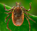
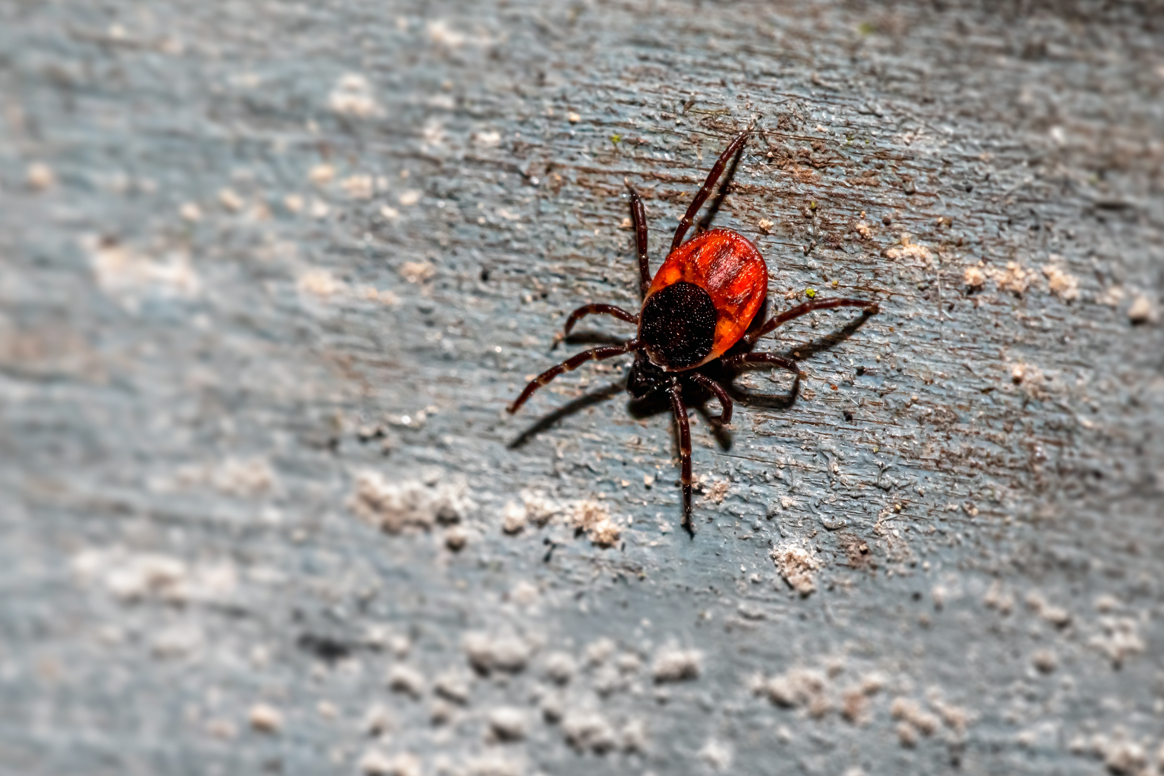
- Blacklegged tick (Ixodes scapularis)[4]
- Brown dog tick (Rhipicephalus sanguineus sensu lato)
- Gulf Coast tick (Amblyomma maculatum)
- Lone star tick (Amblyomma americanum)[5]
- Rocky Mountain wood tick (Dermacentor andersoni)
- Western blacklegged tick (Ixodes pacificus)
Indications
Tick removal is indicated when a patient presents with a tick attached to their body. Indications for treatment beyond removal are based partially on geography.
Individuals should perform a full-body inspection after being in an area of possible tick infestation. Particular attention should be paid to body crevices, areas with hair, and children. If an attached tick is found, identify it first, if possible; the tick might not be intact after removal. Estimate the time of attachment. If the tick has taken blood, a time-dependent process, prophylaxis against tick-borne disease may be indicated in areas of high prevalence.[6]
The American dog tick (D variabilis and D similis), sometimes called the wood tick, transmits tularemia and Rocky Mountain spotted fever. The highest risk of being bitten by American dog ticks occurs in the spring and summer; adult females are the most likely to bite people. D variabilis is widely distributed east of the Rocky Mountains and in limited areas along the Pacific Coast.[3] A newly described D similis is found west of the Rocky Mountains. Further research is required to comprehensively understand the role these species play in disease transmission. [CDC, Regions Where Ticks Live]
The blacklegged tick (I scapularis) is widely distributed in the northeastern upper midwestern United States.[7][8][CDC, Regions Where Ticks Live] I scapularis is a vector of human pathogens, including those causing Lyme disease, anaplasmosis, babesiosis, Borrelia miyamotoi disease, Powassan virus disease, and ehrlichiosis.[9] Humans are at the greatest risk of being bitten in the spring, summer, and fall; bites may occur when temperatures are above-freezing in the winter. Nymphs and adult females are the most likely to bite humans. [CDC, Regions Where Ticks Live] Burtis et al observed that nymphs in Minnesota were most active from May through August, with the peak activity in June; activity did continue through October. Observed nymphal peak activity was directly correlated with the peak incidence of Lyme disease and anaplasmosis cases in that area.[8]
Brown dog ticks (R sanguineus) are the most widespread ticks globally and are well-recognized vectors of many pathogens affecting dogs and occasionally humans.[10][11] At least two lineages of R sanguineus are present in the United States.[12] Dogs are the primary host for the brown dog tick in each life stage, but the tick may also bite humans or other mammals. Brown dog ticks are vectors of Rickettsia rickettsii that cause Rocky Mountain spotted fever from the southwestern United States into northeastern Mexico.[13][14] The brown dog tick is a vector of Rickettsia conorii that causes Meditteranean spotted fever in the Meditteranean basin.[15]
The Gulf Coast tick (A maculatum) is found in coastal areas of the United States, specifically the Atlantic Coast and the Gulf of Mexico. This tick is the primary vector of pathogenic bacteria that cause Rickettsia parkeri rickettsiosis, a form of spotted fever.[16] Larvae and nymphs feed on birds and small rodents, while adult ticks feed on deer and other wildlife. [CDC, Regions Where Ticks Live] However, adult Gulf Coast ticks have been known to transmit R parkeri to humans.[17]
The lone star tick (A americanum) is widely distributed in the southeastern, south-central, eastern, and upper-midwest regions of the United States.[5] Lone star ticks are associated with bacterial, viral, and protozoal pathogens and the newly recognized alpha-gal syndrome, an allergy to red meat and certain mammalian products.[18] This tick transmits two etiologic pathogens of human ehrlichiosis, Ehrlichia chaffeensis and Ehrlichia ewingii, tularemia, and induces southern tick-associated rash illness (STARI). This very aggressive tick will bite humans, and its saliva can be irritating. Redness and discomfort occur at the bite site but may not indicate infection. Adult females have a white dot or “lone star” on their backs. The nymph and adult females most frequently bite humans and transmit pathogens. [CDC, Regions Where Ticks Live]
The Rocky Mountain wood tick (D andersoni) is found in the Rocky Mountain states and southwestern Canada from elevations of 4000 ft to 10,500 ft. The Rocky Mountain wood tick can transmit pathogens, including the Colorado tick fever virus, R rickettsii, and Francisella tularensis, which causes tularemia; it can cause tick-borne paralysis.[19] Adult ticks feed primarily on large mammals and are vectors of human pathogens; larvae and nymphs feed on small rodents. [CDC, Regions Where Ticks Live]
The Western blacklegged tick (I pacificus) is found along the United States Pacific coast, Nevada, Utah, and Arizona. [CDC, Tick Surveillance] I pacificus transmits several human pathogens, including Borrelia burgdorferi, a causative agent of Lyme disease; Anaplasma phagocytophilum, which causes anaplasmosis; Bartonella, and Rickettsiales. Various host species, including small mammals, birds, livestock, and domestic animals, sustain these pathogens within the environment.[20] Larvae and nymphs often feed on lizards, birds, and rodents; adults usually feed on deer. Nymphs and adult females are more frequently reported to bite humans than other life stages of these ticks.[CDC, Regions Where Ticks Live]
Contraindications
There are no contraindications to tick removal.
Equipment
While proprietary tick removal kits are available, tick removal can be accomplished with the following basic equipment:
- Fine-tipped properly aligned sterile tweezers or hemostats
- Alcohol prep wipes
- 2 x 2 in gauze
- Topical antibiotic ointment
- Wound dressing
Personnel
Some individuals can perform this procedure at home. If the patient presents to the clinic, any clinician can perform a tick removal. The clinician is often assisted by a nurse, technician, or medical assistant.
Preparation
The patient should be positioned comfortably with the tick fully exposed. The skin surrounding the attached tick may be cleaned with an alcohol prep pad.
Technique or Treatment
Using the tweezers or hemostat, grasp the tick at the head, directly where it has attached to the skin. Be sure to grasp the head of the tick and not the body. Removing only the body of the tick leaves the head attached to the skin; the head can serve as a nidus for infection and is much more difficult to remove after disembodiment.[21]
Grasp the head of the tick firmly. Then, pull suddenly and directly away from the skin, avoiding twisting or jerking movements. Expect a tiny patch of the skin to come away with the tick. Once the tick has been identified, dispose of it securely.
Post-removal care includes routine cleaning of the skin and the application of a topical antibiotic. Bacitracin is recommended. Apply a wound dressing over the area.
Tetanus prophylaxis is usually not indicated.[22]
Complications
While tick removal is generally straightforward, complications can occur.
Incomplete tick removal leaves residual mouthparts embedded in the skin. These mouthparts can cause local inflammation and serve as a nidus for infection. Signs of infection include erythema, warmth, swelling, tenderness, and purulent discharge.
Tick saliva is a known allergen. Localized and systemic reactions, albeit rare, may occur after tick removal. While a certain amount of erythema surrounding the bite site is usually expected, expanding erythema, wheal formation, urticarial reactions, and dyspnea are more indicative of an allergic reaction.
While not a complication of tick removal, tick-borne infections due to bacteria, viruses, and protozoa transmitted to humans during feeding can and do occur. The risk of disease transmission varies according to the species of tick and the geographical location.[23]
Preventing tick bites to avoid complications includes avoiding tick-infested areas, applying diethyltoluamide (DEET) to the skin as directed on the product label, applying permethrin to clothing, and wearing clothing in a "downward cascade" configuration. For example, wearing pants that cover boot tops ensures that when ticks fall off, they do not lodge in boots.[24]
Clinical Significance
Antibiotic Prophylaxsis
Prophylaxis against Lyme disease following a bite from I scapularis is a chief concern. However, even if the bite occurs in an area where B burgdorferi exists, if the tick has not eaten a blood meal, infection is unlikely. Prophylaxis against Lyme disease is recommended when all of the following circumstances exist:
- the attached tick can be reliably identified as an adult or nymphal I scapularis tick
- the estimated time of attachment is greater than 36 hours based on the degree of engorgement of the tick with blood or certainty about the time of tick exposure
- antibiotic prophylaxis can be initiated within 72 hours of tick removal
- ecologic information indicates that the local rate of infection of ticks with B burgdorferi is greater than 20%
- doxycycline is not contraindicated
If prophylaxis is advisable, a single dose of doxycycline may be offered. There is limited data on the efficacy of chemoprophylaxis initiated more than 72 hours following tick removal. More than 20% of the I scapularis population is infected with B burgdorferi in parts of New England, the mid-Atlantic States, Minnesota, and Wisconsin, but not in most other locations in the United States. Whether antibiotic prophylaxis after a tick bite will reduce the incidence of anaplasmosis or babesiosis is unknown.[25]
It is not recommended to use amoxicillin instead of doxycycline in persons for whom doxycycline is contraindicated because of the absence of data on an effective short-course regimen for prophylaxis, the likely need for a multi-day regimen (and its associated adverse effects), the efficacy of antibiotic treatment of Lyme disease if infection were to develop, and the low risk that a person with a tick bite will develop a serious complication of Lyme disease.[6] Patients should be educated to seek care if they develop signs or symptoms of Lyme disease after tick removal, whether or not they are given prophylaxis.
Enhancing Healthcare Team Outcomes
In most parts of the United States, the highest incidence of tick bites occurs during spring and summer. Patients may present to the primary care clinic, urgent care, or emergency department with an attached tick. All clinicians should know how to manage ticks. In some geographical regions, prophylactic antibiotics may be considered due to a high incidence of tick-related disease. The interprofessional team may consider an infectious disease consultation to determine what if any, type of treatment is needed after tick removal. Patients with tick bites require education regarding the potential complications and disease transmission. The pharmacist should ensure medication compliance if a systemic infection is being treated. If noncompliance is a concern, the clinical team should be contacted.[26]