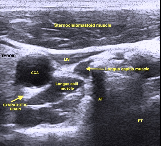
[1]
McLean B. Safety and Patient Acceptability of Stellate Ganglion Blockade as a Treatment Adjunct for Combat-Related Post-Traumatic Stress Disorder: A Quality Assurance Initiative. Cureus. 2015 Sep 10:7(9):e320. doi: 10.7759/cureus.320. Epub 2015 Sep 10
[PubMed PMID: 26487996]
Level 2 (mid-level) evidence
[2]
Wang D. Image Guidance Technologies for Interventional Pain Procedures: Ultrasound, Fluoroscopy, and CT. Current pain and headache reports. 2018 Jan 26:22(1):6. doi: 10.1007/s11916-018-0660-1. Epub 2018 Jan 26
[PubMed PMID: 29374352]
[3]
Duong S, Bravo D, Todd KJ, Finlayson RJ, Tran Q. Treatment of complex regional pain syndrome: an updated systematic review and narrative synthesis. Canadian journal of anaesthesia = Journal canadien d'anesthesie. 2018 Jun:65(6):658-684. doi: 10.1007/s12630-018-1091-5. Epub 2018 Feb 28
[PubMed PMID: 29492826]
Level 1 (high-level) evidence
[4]
Gunduz OH, Kenis-Coskun O. Ganglion blocks as a treatment of pain: current perspectives. Journal of pain research. 2017:10():2815-2826. doi: 10.2147/JPR.S134775. Epub 2017 Dec 14
[PubMed PMID: 29276402]
Level 3 (low-level) evidence
[5]
Datta R, Agrawal J, Sharma A, Rathore VS, Datta S. A study of the efficacy of stellate ganglion blocks in complex regional pain syndromes of the upper body. Journal of anaesthesiology, clinical pharmacology. 2017 Oct-Dec:33(4):534-540. doi: 10.4103/joacp.JOACP_326_16. Epub
[PubMed PMID: 29416250]
[6]
Narouze S. Ultrasound-guided stellate ganglion block: safety and efficacy. Current pain and headache reports. 2014 Jun:18(6):424. doi: 10.1007/s11916-014-0424-5. Epub
[PubMed PMID: 24760493]
[7]
Imani F, Hemati K, Rahimzadeh P, Kazemi MR, Hejazian K. Effectiveness of Stellate Ganglion Block Under Fuoroscopy or Ultrasound Guidance in Upper Extremity CRPS. Journal of clinical and diagnostic research : JCDR. 2016 Jan:10(1):UC09-12. doi: 10.7860/JCDR/2016/14476.7035. Epub 2016 Jan 1
[PubMed PMID: 26894152]
[8]
Chang KV, Lin CP, Hung CY, Özçakar L, Wang TG, Chen WS. Sonographic Nerve Tracking in the Cervical Region: A Pictorial Essay and Video Demonstration. American journal of physical medicine & rehabilitation. 2016 Nov:95(11):862-870
[PubMed PMID: 27362696]
[9]
Bhatia A, Flamer D, Peng PW. Evaluation of sonoanatomy relevant to performing stellate ganglion blocks using anterior and lateral simulated approaches: an observational study. Canadian journal of anaesthesia = Journal canadien d'anesthesie. 2012 Nov:59(11):1040-7. doi: 10.1007/s12630-012-9779-4. Epub 2012 Sep 6
[PubMed PMID: 22956268]
Level 2 (mid-level) evidence
[10]
Chang KV, Wu WT, Özçakar L. Ultrasound-Guided Interventions of the Cervical Spine and Nerves. Physical medicine and rehabilitation clinics of North America. 2018 Feb:29(1):93-103. doi: 10.1016/j.pmr.2017.08.008. Epub 2017 Oct 7
[PubMed PMID: 29173667]
[11]
Peterson K, Bourne D, Anderson J, Mackey K, Helfand M. Evidence Brief: Effectiveness of Stellate Ganglion Block for Treatment of Posttraumatic Stress Disorder (PTSD). 2017 Feb:():
[PubMed PMID: 28742302]
[12]
Wei K, Feldmann RE Jr, Brascher AK, Benrath J. Ultrasound-guided stellate ganglion blocks combined with pharmacological and occupational therapy in Complex Regional Pain Syndrome (CRPS): a pilot case series ad interim. Pain medicine (Malden, Mass.). 2014 Dec:15(12):2120-7. doi: 10.1111/pme.12473. Epub
[PubMed PMID: 25537318]
Level 2 (mid-level) evidence
[13]
Mulvaney SW, Lynch JH, Hickey MJ, Rahman-Rawlins T, Schroeder M, Kane S, Lipov E. Stellate ganglion block used to treat symptoms associated with combat-related post-traumatic stress disorder: a case series of 166 patients. Military medicine. 2014 Oct:179(10):1133-40. doi: 10.7205/MILMED-D-14-00151. Epub
[PubMed PMID: 25269132]
Level 2 (mid-level) evidence

