Continuing Education Activity
Ventral hernias of the abdomen are non-inguinal, non-hiatal defects in the fascia of the abdominal wall. They are commonly seen in clinical practice. The repair of these abdominal wall defects is a common surgery performed by general surgeons. This activity describes the pathophysiology, evaluation, and management of ventral hernias and highlights the role of the interprofessional team in caring for affected patients.
Objectives:
- Identify the etiology of ventral hernias.
- Describe the presentation of a patient with a ventral hernia.
- Outline the treatment and management options available for ventral hernias.
- Explain interprofessional team strategies for improving care coordination and communication to advance the management of ventral hernias and improve outcomes.
Introduction
Ventral hernias of the abdomen are defined as a non-inguinal, nonhiatal defect in the fascia of the abdominal wall. Annually, there are about 350,000 ventral hernia operations. The repair of these abdominal wall defects is a common surgery performed by general surgeons. Surgery is typically recommended for individuals with acceptable operative risk, symptomatic hernias, or those at elevated risk of developing complications from a hernia. They can affect an individual’s quality of life and can lead to hospitalizations and even death in some cases.[1][2][3]
Etiology
Etiologies of a ventral hernia can be broken down into 2 main categories; acquired or congenital. The vast majority of hernias that general surgeons see and treat are acquired; however, some individuals live with their ventral hernias from birth for prolonged periods of time before having them surgically repaired. Common causes of acquired ventral hernias include previous surgery causing an incisional hernia, trauma, and repetitive stress on naturally weak points of the abdominal wall. These naturally occurring weak points in the abdominal wall include the umbilicus, semilunar line, ostomy sites, bilateral inguinal regions, and esophageal hiatus. Obesity is a large component of hernias as well because it stretches the fascia of the abdomen causing it to weaken. Specifically, the action of repetitive weight gain and loss leads to weakening.[4]
Epidemiology
In 2006, 348,000 ventral hernia repairs were performed in the United States, and it was estimated to cost approximately $3.2 billion. This is a large burden on the healthcare system with the majority of the cost coming from emergency repairs or complications postsurgically. In the post-operative setting, patients have an approximately 10% risk of developing a hernia following a midline laparotomy, 5% following a transverse muscle splitting incision, and less than 1% following laparoscopic repair.[5][6]
Pathophysiology
The anterior abdominal wall is made of many layers including skin, fat, fascia, muscle, and peritoneum. The order of the layers change depending on the location you enter the abdomen perpendicularly. A point approximately midway between the umbilicus and pubic symphysis is an imaginary line called the arcuate line. At this point, the layers of the abdomen, with respect to the rectus, change in orientation. Above the arcuate line, the fascia of the internal oblique aponeurosis envelops the rectus muscle. The external oblique aponeurosis always lays anterior to the internal oblique aponeurosis and the transversus abdominis aponeurosis always posterior to it. However, below the arcuate, line all 3 layers of aponeurosis become anterior to the rectus muscle, and it is no longer enveloped. Instead, the only fascial layer below the rectus is the transversalis fascia which is separate from the transversus abdominis aponeurosis.[7][8]
Repetitive stresses on the abdominal wall from increased intra-abdominal pressure lead to microscopic tears of tissue. Over time this can decrease the strength of tissue, predisposing individuals to hernia formation. Several instances cause increased intra-abdominal pressures that place individuals at increased risk including constipation, physical labor, childbirth, excessive coughing from lung disease or even frequent vomiting from diseases like bulimia nervosa.
Tissue strength following surgery can only achieve an 80% tensile strength of the previous maximum. This effect is additive as well, so after a second midline laparotomy, the maximum tissue strength would be 80% of 80%, which is 64%. This 80% predicted tensile strength is under perfect conditions as well assuming no evidence of malnutrition or infectious complications.
Histopathology
It is common practice to send the hernia sac from ventral wall hernias. A large retrospective review showed that 7 of 576 ventral wall hernias revealed a malignancy. Five of these lesions were not seen on examination. Other pathologies that were revealed included appendicitis, endometriosis, a perivascular epithelioid cell tumor, and pseudomyxoma peritonei. The review led to the conclusion that ventral hernias should be submitted for histologic evaluation.
History and Physical
The presentation of an abdominal wall hernia is usually pain, swelling or fullness at the site of occurrence that can change with position or Valsalva. In some cases when a hernia is incarcerated or strangulated, the enlargement may be erythematous or cause an asymmetry. In most cases, the diagnosis of an abdominal hernia can be made by history and physical exam but severe obesity, which is a major risk factor, can limit the exam. It is very important that during the exam the patient is examined in multiple positions as hernias can change with exertion or even standing.
There are additional questions that should be explicitly asked of patients presenting with ventral hernias. Beginning with the history of present illness, details surrounding the hernia should include the first time they noticed it, any inciting events, associated pain, erythema, constipation, nausea or vomiting, size of the bulge, change in size, what causes the change in size, history of previous hernias, weight change and a detailed surgical/medical history. Important social questions including the patient's occupation, dietary habits, exercise habits, smoking history, and alcohol consumption should also be included in the history. Patients should be asked specifically about the family history of a connective tissue disorder as this is typically hereditary and can lead to hernia formation.
Evaluation
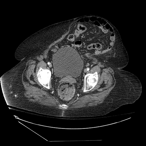
Multiple adjunct examinations can be performed to assist with diagnoses including ultrasound, CT scan or MRI. If the diagnosis is in question or even the size of the hernia defect in unclear these studies can be ordered to aid in diagnosis or preoperative planning. Preoperative medical clearance is another important aspect in operative planning. Each surgeon should follow a set of guidelines for this and assume the need for a general anesthetic. It is possible to do open ventral hernia repairs without general sedation; however, it is unlikely the patient will be completely relaxed, and therefore, it makes the operation more challenging with a possibly inferior result. The risk of ventral hernia repair varies greatly from low risk with a small umbilical hernia to a major risk with a large component separation. Patients with lung disease of a prolonged history of smoking would benefit from pulmonary function tests pre-operatively. It is also important to make sure that any screening exams are performed before abdominal surgery, it would be unfortunate to have to perform a colectomy for colon cancer on a patient 6 months after doing a ventral hernia repair. There should be strict adherence to discontinuation of antiplatelet and anticoagulation medications secondary to hematoma formations increasing the probability of infectious complications.[9][10]
Treatment / Management
The most common treatment of ventral hernias includes surgery. Asymptomatic hernias are repaired on an elective basis, but those presenting with strangulation require immediate surgery. Incarceration without strangulation is not a surgical emergency; however, the risks and benefits of surgery should be discussed with the patient, and a patient with reasonable operative risk should have their hernia repaired within a sensible time frame. Non-surgical management of abdominal wall hernias with the use of binders, trusses, or corsets is not considered to be effective. However, this may be the only option in a patient who is not a reasonable candidate for surgery. [11][12][13]
Over the years, many types of surgical techniques have been developed to repair hernias. There are many tenants of hernia repair. The most important being a tension-free closure, but others include the use of a mesh with 3 to 5 cm of overlap, meticulous handling of the mesh, preventing surgical site infections, and using a sublay technique with the closure of the fascia if possible. The most basic approach is a primary open repair without mesh, which should typically be reserved for defects in the fascia of less than 2 cm. An open repair with mesh has several options including what type of mesh and where to place the mesh.
Laparoscopic ventral hernia repair when compared against open techniques has consistently showed decreased overall complication rates, decreased hospital length of stay, and a quicker return to work. Although it has not been consistently statistically significant, a large portion of available literature shows the recurrence rates are slightly lower in laparoscopic repair. The disadvantages of laparoscopy include a higher potential for visceral injury and it is technically more difficult. There has been development of wristed laparoscopic instrument that give additional freedom of motion during operating but additional research is needed to detect significant benefit.
Robotic ventral hernia repairs have also become popular secondary to increased freedom of motion during surgery. Closing the fascial defect robotically is far easier from a technical standpoint than attempting it with classical laparoscopic instruments. The benefits of laparoscopy are retained secondary to the smaller incisions that can be maintained. Robotic surgery is typically more expensive and has longer operative times than laparoscopy, and at this point, no landmark trials have demonstrated superiority of robotic surgery in comparison to laparoscopy.
Component separations can be performed several different ways and are typically reserved for large defects in which a tension-free closure cannot be achieved. All of the techniques require adhesiolysis with reduction of the hernia and typical mesh placement. The open technique with an onlay mesh consists of developing large skin flaps, about 5 cm beyond the midline, exposing the lateral portions of the rectus. The external oblique is then incised 2 cm lateral to linea semilunaris and extended superiorly and inferiorly while separating it from the internal oblique. This allows medialization of the rectus muscle and closure of the defect. Mesh is then used to reinforce the closure in an onlay fashion. It is estimated to allow for tension-free closure of defects up to 10 cm in diameter. During the endoscopic component separation the hernia is reduced after adhesiolysis and incisions are made laterally to the rectus. The external oblique fascia is incised and the muscle is split all the way to the posterior fascia. A balloon is then inserted along the posterior fascia and inflated underneath of the external oblique muscle down the level of the anterior superior iliac spine creating a large space. Using additional ports the external oblique can then be incised to allow medialization of the rectus and repair of the hernia.
Transversus abdominis muscle release (TAR), also known as a posterior component separation, is another option for large hernias and consists of developing the retro-muscular space from the medial rectus into the space between the transversus abdominis and internal oblique. After the posterior rectus sheath is released, it is incised laterally, and the transversus abdominis is released medial to the linea semilunaris to expose a broad plane that extends from the central tendon of the diaphragm superiorly, to the space of Retzius inferiorly, and laterally to the retro-peritoneum on both sides. This preserves the neurovascular bundles innervating the medial abdominal wall. The mesh is placed in a sublay fashion above the posterior fascial layer but below the rectus and internal oblique muscles. The posterior rectus fascia then is advanced medially and closed while the linea alba is restored anterior to the mesh. These cases can be very long cases and technically very challenging.
A different type of a hernia that can affect the abdominal wall is a parastomal hernia. It is estimated that up to 30% of patients with ostomies can develop parastomal hernias. Some types of ostomies are at higher risk than others. Loop colostomies are at the highest risk followed by end colostomies, loop ileostomies, and end ileostomies. Currently, the only strategy to prevent hernia formation is the use of a prophylactic mesh when the ostomy is created. Patients that are planning an ostomy reversal typically have the repair delayed until the reversal of the ostomy, but sometimes the repair can be an emergency. There are multiple types of repairs, and the 2 that will be discussed here include the modified Sugarbaker and keyhole techniques. Both procedures can be performed laparoscopically or open. After adhesiolysis, the modified Sugarbaker technique consists of lateralizing the bowel by tracking the bowel from the hernia sac between the abdominal wall and the prosthesis into the peritoneal cavity. Essentially putting a patch over the defect and having the bowel enter the abdominal cavity laterally to the mesh. The keyhole technique is performed by making a slit in the mesh and fitting the mesh around the bowel before fixating the mesh, thereby patching the defect. In some studies the Sugarbaker technique had a lower incidence of recurrence but larger studies are needed to achieve statistical significance.
Mesh can be divided into synthetic or biologic. The decision of which mesh to use is mainly up to the surgeon, but there are instances where one should be used over the other. Most synthetic prosthetic grafts can be categorized as derived from polypropylene, polyester, or polytetrafluoroethylene (PTFE). In the late 1990s, lightweight mesh came in the market and has now been widely accepted as a superior mesh, but it still can become infected and have recurrences. The size of the pores in the mesh has also come under scrutiny and they have discovered there is a major benefit to macro-porous mesh, defined as pores larger than 3 mm. These cause fewer incidences of infection, and if the do become infected, there is a much higher chance that treatment with antibiotics can prevent explantation. Mesh composed of ePTFE has a good profile for adhesion risk but a high risk of infection. In contrast, polypropylene mesh has a lower infection risk but little flexibility and a high adhesion risk. There is also 2-sided mesh designed for intra-peritoneal placement in which one side is coated with an anti-adhesive barrier. This is not 100% successful, but studies have shown decreased adhesion formations and easier adhesiolysis of those that do form. There is also sutureless mesh that sticks to the tissues designed to reduce post-operative pain and prevent mesh migration. Absorbable mesh-like vicryl can be used in infected fields, but it will absorb over time leaving only native tissue.
The number and type of biologic grafts has expanded greatly over the last decade. The huge downside to biologic meshes is they are typically very expensive. They are usually reserved for infected or contaminated fields and the strength of the repair is considered inferior to synthetic mesh placement. They typically consist of an acellular collagen matrix derived from human dermis or porcine small intestine submucosa. These biologic meshes still generate a foreign body reaction as well so they can cause adhesions but are unlikely to become infected.
Differential Diagnosis
The differential diagnosis of a hernia should be a relatively short list. Additional pathologies would include diastasis recti, abscess, muscle strain, seroma, wound hematoma, lymphadenopathy, soft tissue malignancy, and rectus sheath hematomas.
Staging
Unfortunately, there is not currently a universal classification system for ventral hernias. One of the more accepted classification systems is the European Hernia Society (EHS) classification system. They separated the system into primary abdominal wall hernias and incisional abdominal wall hernias. A primary ventral hernia that not associated with a previous operation is usually in a limited number of locations, subdivided into midline and lateral, while the classification can be limited to 2 variables: length and width. The classifications of incisional abdominal wall hernias are more complicated as they can occur anywhere on the abdomen, but again, they are documented in terms of length and width. The limitation of this system is that it does not include individual patients risk factors and wound classification. However, a classification complex enough to encompass all of the important variables would be difficult to remember and unlikely to be embraced by the surgical community. In instances where a significant amount of the visceral contents are in the hernia sac, some sources define this as half of the abdominal contents; it is considered to be a ventral hernia with loss of domain.
Prognosis
Patients have varying prognoses after ventral hernia repair. The circumstances surrounding the original operation have the highest predictive value of post-operative complications. Emergency operations for strangulation that require bowel resection are associated with higher morbidity and recurrence secondary to the face at a minimum the case has become cleanly contaminated, and synthetic mesh should not be used. Wound class is an important variable in determining the risk of morbidity from an operation and should be documented in the operative record. A clean wound (class I) is an incision where no inflammation is encountered, there was no break in sterile technique, and the respiratory, alimentary and genitourinary tracts were not entered. A clean-contaminated wound (class II) is an incision where the respiratory, alimentary, or genitourinary tract is entered under controlled conditions but with no contamination encountered. A contaminated wound (class III) is an incision where there could have been a major break in sterile technique, obvious spillage from the gastrointestinal tract, or an incision in which acute, nonpurulent inflammation is encountered. Open traumatic wounds that are more than 12 to 24 hours old also fall into this category. A dirty or infected would (class IV) is an incision where the viscera may have been perforated, acute inflammation with pus is encountered during the operation and for traumatic wounds where treatment is delayed, or there is fecal contamination/devitalized tissue present.[14]
Complications
Recurrence of a ventral hernia after the repair has varying rates over time, but with the introduction of mesh, the recurrence rates have dropped significantly. Recurrence rates differ among the type of repairs: in laparoscopic repairs with mesh are around 10% to 12%, open-mesh repair 13% to 15%, and open-tissue repair 18% to 20%. Component separation estimates recurrence rates as high as 20% in large studies, but this is not comparable with other operations secondary to large defects and need for reconstruction of the abdominal wall.
Mesh infection is a catastrophic complication of ventral hernia repairs because it is typically followed by a second operation that is more complex and associated with a high chance for recurrence of a hernia. There are many risk factors including a high body mass index (BMI), chronic obstructive pulmonary disease (COPD), abdominal aortic aneurysm repair, prior surgical site infection, mesh type, longer operative time, lack of tissue coverage of the mesh, enterotomy, and surgical site infections. With a mesh infection, it is much more common to require explantation of the mesh, but salvage is a possibility with antibiotics.
Respiratory morbidity following ventral hernia repair is specifically a concern of abdominal wall reconstruction secondary to decreasing the volume of the abdominal cavity. This exerts upward pressure on the diaphragm and can lead to hypoxia and intubation. There are institutions with protocols to prevent morbidity after these demanding surgeries. Evidenced-based literature to prevent respiratory morbidity includes sufficient pain control using PCA, regional blocks or epidurals, early ambulation after surgery, and not routinely using nasogastric tubes. Evidence does not support the use of lung expansion therapies such as deep breathing exercises, incentive spirometry, and CPAP, but they are still commonly used.
Postoperative and Rehabilitation Care
Patients should limit themselves to a 10-pound (4.5 kg) lifting restriction within the first week, 20 pounds (9 kg) for the second week, and slowly advance to full activity over a period of 6 weeks. It is important to limit the narcotics during the postoperative phase secondary to addiction potential and also to prevent constipation. Multi-modal pain control with acetaminophen, anti-inflammatory, neuropathic, and muscle-relaxing medication, in addition to narcotics, have been shown to decrease opiate usage. Additional adjuncts including regional blocks, long-acting, local anesthetics, as well as post-surgical nerve blocks have also been successful in decreasing opiate usage. Stool softeners and laxatives in the postoperative phase are common practice to prevent straining and bloating. There are typically no dietary restrictions, but patients should eat a high fiber diet in the postoperative period. Patients can shower in the 24 to 48 hours window following surgery. It is useful to provide patients with postoperative instructions at multiple points throughout their preoperative and postoperative course. Offering literature on the topics is useful as well. Patient education has been shown in multiple specialties to help prevent postoperative complications.
Deterrence and Patient Education
Prevention of acquired ventral hernias is difficult because it is mainly focused on prevention of incisional hernias. Enforcing or monitoring whether patients adhere to the instructions is impossible. Meticulous closure of the abdomen should be performed with a suture length to wound length ratio of greater than 4. Also, each bite of fascial closure should travel longitudinally along the incision and have a bite depth between 5 and 10 mm. It is very important that during the procedure, surgeons obtain a tension-free closure and close all port site 10 mm or larger. Incisional hernias have been documented to occur in port sites smaller than 10 mm, but these are rare. The closure of larger abdominal incisions also needs further studies to define the best suture for closure. However, there have been many evidence-based papers that recommend a running slowly absorbable or non-absorbable monofilament sutures for incision closure. The non-absorbable suture has achieved the lowest rates for hernia formation but at an increased risk of chronic pain and sinus drainage from fistula to the suture. The benefit of monofilament suture is secondary to a decreased infection risk when compared with multi-filament suture.
The most important aspect of preventing a ventral hernia is to prevent a wound infection in a surgical setting. Wound infections increase the chance of hernia development by a statistically significant amount. It is also associated with higher mesh infections that have a higher likelihood of requiring a second surgery. Laparoscopic repairs have been consistently linked with lower wound infections than open repairs. Patient optimization prior to surgery has also consistently been linked to lower rates of infections. Encouraging smoking cessation, improved nutrition, weight loss, physical rehabilitation, and tight glycemic control helps, as well as overall patient selection. There are also many preoperative and intraoperative measures to decrease surgical site infection, for example, preoperative antibiotics, prevention of hypothermia, sterile technique, skin preparation and many others. The use of abdominal binders has not been shown to decrease the incidence of ventral hernia occurrence or recurrence. The only consistent benefit of abdominal binders has been the patients subjectively feel more comfortable.
Pearls and Other Issues
Tension-free repair with mesh for ventral hernias over 2 cm in size is the standard of care. A retro-rectus mesh placement in the pre-peritoneal space has become the optimal placement for the mesh. Loss of abdominal wall domain requires a complex operation with high morbidity to achieve a tension-free repair. Port sites 10 mm and higher should have fascial closures; some port site hernia can occur at smaller sizes.
Enhancing Healthcare Team Outcomes
Ventral hernias are very common and do present a challenge because of the risk of recurrence. Besides the surgeon, these hernias are ideally managed by a dietitian, nurse, and a physical therapist. Patient education has been a very popular topic in literature and hospital settings. Giving patients literature and discussing outcomes with instructions on multiple aspects throughout their encounters has shown to be very effective. Also providing patients with available resources to ask questions or contact a healthcare provider has been shown to decrease returns to the emergency room within 30 days.
The outcome of ventral hernias depends on the size and other patient comorbidity. Elective hernia repair has the best outcomes, but for patients with incarceration or strangulation, the outcomes are guarded. Mortality rates in excess of 5% have been reported in presence of a strangulated hernia. While laparoscopic herniorrhaphy has led to relatively easy repair, the procedure has been known to be associated with serious complications that have been related to bowel injury from the instrumentation.[15][16][17]