Continuing Education Activity
Lower blepharoplasty is performed for many reasons, although the commonest presentation by patients is one of "looking old" or "looking tired." Other complaints may include "baggy eyelids," swollen eyelids that are worse in the morning, and "my photographs make me look tired." There are many different techniques available when performing lower blepharoplasty, and modern surgeons must perform a careful preoperative assessment in order to choose procedures to optimize cosmetic and functional results for each patient. Surgeons must possess a thorough understanding of each surgical technique to ensure adequate patient selection for the procedure and ensure optimal outcomes. This activity reviews the role of the interprofessional team in the care of these patients.
Objectives:
- Identify the anatomical structures, indications, and contraindications of lower lid blepharoplasty via a subciliary approach.
- Describe the technique necessary to perform blepharoplasty via a subciliary approach.
- Review the potential complications of blepharoplasty via a subciliary approach.
- Outline interprofessional team strategies for improving care coordination and communication when considering blepharoplasty via a subciliary approach to improving outcomes.
Introduction
Lower blepharoplasty is performed for many reasons, although the commonest presentations include a feeling of "looking old" or "looking tired." Other complaints may include "baggy eyelids," "swollen eyelids, worse in the morning," and "my photographs make me look tired." Some surgeons have noticed an increase in facial cosmetic consultations they attribute to the omnipresence of social media.[1]
Although there are many different techniques available when performing lower blepharoplasty, modern surgeons have come to appreciate that preoperative assessment allows a combination of procedures to give the best cosmetic and functional results. It is now expected that the surgeon will be familiar with a variety of techniques and can combine them to tailor such techniques to each patient, rather than always performing the same procedure on every patient.
Blepharoplasty can be defined as "changing the shape of the eyelid" and can be performed for functional and aesthetic indications.
Subciliary blepharoplasty is the approach to the deeper lower eyelid structures via a transcutaneous incision placed in the subciliary crease and can be combined with a lateral canthotomy and cantholysis for lid-tightening if required. Some surgeons will use this approach when repairing the orbital floor or zygomatic-maxillary complex (ZMC) fractures.
Transcutaneous blepharoplasty is usually indicated in cases with skin and muscle laxity, with or without fat prolapse. Traditionally, the skin is incised 1 mm below the lash line in the subciliary area. Once the skin is incised, there are two possible variations that are named depending upon the plane of dissection. A 'skin flap' can be raised, elevating the thin eyelid skin off of the orbicularis oculi muscle. In 1951, Castanares described the 'skin-flap' technique as best suited for eyelids with excess lax skin and atonic orbicularis muscle.[2] The second technique is termed the 'skin-muscle' flap, where the orbicularis oculi muscle is incised as it attaches to the tarsal plate, and the plane of dissection is deep to the orbicularis and superficial to the orbital septum. Mcindoe-Beare popularized this technique and proposed its use in younger patients with robust orbicularis muscle tone.[3]
Regardless of the specific plane of dissection used, lower lid malpositions, particularly ectropion, may be encountered in the subciliary approach owing to contractile scarring that everts the lash line and grey line away from the globe. The most common causes of this complication include unaddressed eyelid laxity, overzealous skin excision, denervation of the orbicularis, breach of the orbital septum, or unfavorable scarring. A meticulous preoperative examination can identify eyelid laxity, which must then be addressed at the time of blepharoplasty. In an effort to avoid the potential for ectropion, Bourget introduced the transconjunctival approach, which was popularized by Zarem and Resnik as beneficial in avoiding ectropion.[4] This approach is discussed in an additional article on StatPearls.com.
The selection of a transcutaneous approach or a transconjunctival approach for lower lid rejuvenation is an area of active controversy with vocal advocates on all sides. It behooves the surgeon to be familiar with both approaches, allowing treatment to be tailored to the patient. Maffi et al. reviewed 2007 patients over a period of 30-years who underwent traditional transcutaneous blepharoplasty without additional support by a senior experienced surgeon and reported only 0.4% symptomatic lid malposition post-surgery, which supports the safety and effectiveness of this procedure.[5] This is a similar, low complication rate to that seen in transconjunctival approaches.[6]
Contemporary lower lid blepharoplasty has also benefitted from advances in the understanding of the anatomical changes of the aging face, the importance of orbicularis retaining ligament (ORL), the orbitomalar sulcus deformity, and tear trough deformity. Current literature focuses on smoothing the lid-cheek junction for a more youthful look using techniques like the release of retaining ligaments, fat transposition, and mid-face augmentations. Also, lid-anchoring procedures have been emphasized to treat any lid laxity. Although these techniques for lid-cheek junction smoothing can be accomplished through the transconjunctival approach, the transcutaneous approach allows the excellent field of exposure for fat transposition, ORL release, midface-lift procedures as well as lateral canthal tightening procedures with the additional advantage of skin redraping.
Many surgeons routinely integrate or combine these approaches to get the best outcomes and minimizing complications in lower lid blepharoplasty. For a patient without excess skin and muscle, transconjunctival blepharoplasty along with fat transposition and skin laser treatment can be performed. Patients with excess skin-muscle require combining skin-muscle flap through subciliary incision with ORL release, fat transposition, lateral canthal support, and minimal removal of fat through the conjunctiva. Similarly, multimodality 5-step blepharoplasty, described by Rohrich et al.[7] includes (1) malar fat augmentation, (2) minimal fat resection transconjunctivally preserving orbicularis, (3) ORL release, (4) lateral canthal tightening procedure, (5) minimal skin removal via subciliary incision.
Anatomy and Physiology
Eyelid Anatomy
Grossly, eyelids are divided into the orbital and tarsal parts by a sulcus or furrow. Additionally, there is the nasojugal fold medially and the malar fold laterally. The skin of the eyelid is the thinnest in the body, below which lies scant subcutaneous tissue. There is a transition to thicker skin from the lower eyelid to the cheek.
Concentrically arranged striated muscle, the orbicularis oculi, lies beneath the skin and subcutaneous tissue. It is divided into three parts, namely, the orbital, preseptal, and pretarsal parts. The orbital part originates from the medial canthal tendon, the orbital process of the frontal bone, and the frontal process of the maxilla, forming a complete ellipse and inserts just below the point of origin. A surgically relevant ligamentous attachment, the orbital retaining ligament (ORL), attaches the orbital portion of the orbicularis to the periosteum of the maxilla[8], giving the lid-cheek demarcation. Similarly, in the area of the tear trough, the orbicularis muscle directly attaches to the orbital rim. The preseptal portion takes origin from the upper and lower border of the medial canthal tendon, inserting onto the lateral palpebral raphe. Likewise, the pretarsal portion originates medially from the posterior lacrimal crest and anterior limb of the medial canthal tendon, inserting onto the lateral canthal tendon. This portion of the pretarsal muscle plays an important role in the drainage of tears and the lacrimal pumping mechanism of blinking. The orbicularis oculi muscle is supplied by the temporal and zygomatic branches of the facial nerve.
The orbital septum, which lies beneath orbicularis oculi, is a thin, fibrous framework that begins anatomically at the arcus marginalis. It acts as a barrier and holds orbital fat in position. In the lower lid, there exist three fat pockets contained within the medial, central, and lateral compartments. The medial fat pad is separated from the central fat by the inferior oblique muscle. The medial fat is paler in appearance as compared to the yellowish lateral fat pad. The central and lateral fat pads are separated by a fascial layer extending off the capsulopalpebral fascia, and removal of this barrier improves access to the lateral fat pad. Inferiorly, the malar fat lies in the subcutaneous plane extending over the inferior orbital rim.
Effects of Aging
Aging skin becomes thin, wrinkled, less elastic, hyperpigmented with actinic changes as well as increased laxity of the orbicularis muscle and canthal tendons.
With aging, there is gradual tissue atrophy, loss of adnexal structural support of the tarsus, canthal tendons, and orbicularis muscle, as well as thinning of the skin and loss of rete ridges, which leads to orbital fat prolapse, ptosis, and eyelid malposition. These changes lead to a more pronounced demarcation of the eyelid from the cheek, resulting in two convexities with a sulcus between them.
The upper convexity is due to the prolapse of orbital fat in the lower lid precipitated by the laxity of the orbital septum and weakening of the suspensory ligament of Lockwood, which in turn leads to downward globe descent and pseudoherniation of fat through an intact but weakened orbital septum.[9]
The lower convexity is a result of the descent of malar fat as well as volume loss, causing the phenomenon known as 'skeletonization of inferior orbital rim,' which refers to the increased visual prominence due to surrounding tissue changes. The inferior displacement of this subcutaneous fat is due to age-related atrophy and gravity.
The sulcus separates the superior bulge from the inferior. The sulcus is referred to as tear trough deformity medially and laterally as the lid-cheek junction. Medially the nasojugal groove, or tear trough, extends from the medial canthus to the mid pupillary line and is due to the dense connection between palpebral orbicularis muscle and maxilla. Similarly, malar sulcus or lid-cheek junction starts from malar prominence and extends until it blends with nasojugal sulcus in the subcutaneous plane. But in the sub-orbicularis plane, the orbicularis muscle is not directly attached to the bone; it is through a ligament (the ORL). Since both the nasojugal and malar grooves have a muscular and facial attachment to the periosteum of the inferior orbital rim, the superior bulge is accentuated at the sulcus. So, the release of these attachments is very important to blend this demarcation and correct the double convexity effectively.
Indications
Lower lid blepharoplasty via a subciliary approach may be performed for a variety of cosmetic and functional indications. Some of the most common include:
1. Dermatochalasis: Excess lower eyelid skin with thinning and laxity forms the most common indication for transcutaneous lower lid blepharoplasty. It can be associated with loss of orbicularis muscle tone or its hypertrophy and canthal laxities, which need to be addressed during surgery.
2. Steatoblepharon: Fat prolapse in the lower lid is another common indication. Many prefer a transconjunctival approach in young patients with no skin excess. Transcutaneous is mainly preferred in cases with fat prolapse and excess skin. The tear trough and lid-cheek junction giving double convexity appearance is another concern of these patients, which can be blended with fat repositioning.
3. Ectropion or entropion: Senile lid changes like ectropion or entropion with associated redundant skin forms a functional indication for transcutaneous lower lid blepharoplasty.
Contraindications
The contraindications for transcutaneous blepharoplasty are similar to those for any purely elective or aesthetic surgery. Most are relative contraindications and can include:
Absolute Contraindications
- Patients with unrealistic goals of surgery
- Only-seeing eye
- Active blepharitis or hordeolum
- Uncontrolled glaucoma
- Active myasthenia gravis
Relative Contraindications
- Significant medical comorbidities rendering surgery unsafe (uncontrolled hypertension, heart failure, pulmonary disease, bleeding dyscrasias, and many others)
- History of hypertrophic scarring or keloid formation
- History of pemphigus vulgaris or Stevens-Johnson syndrome
- Anticoagulant therapy: Should be stopped 1 to 2 weeks prior to surgery
- Pseudoproptosis or proptosis: The conditions such as thyroid orbitopathy can also present with fat prolapse with or without proptosis because of increased orbital volume. These patients may have lid retraction, lid lag, and muscle restriction, causing diplopia. These patients may require orbital decompression instead of blepharoplasty.
Equipment
The ophthalmic plastic surgical set instruments will be required:
- Local anesthesia (2% lignocaine with adrenaline)
- Topical anaesthetic (0.5% proparacaine)
- Surgical marker
- Radio-frequency cautery with fine cutting needles
- Bipolar cautery
- Retractors (Desmarre retractor)
- Tissue forceps (fine-toothed)
- Small scissors (Wescott or Steven scissors)
- Needle holder (Barraquer or Castroveijo)
- Periosteal elevator (Freer)
- Artery forceps (varying sizes/ mosquito forceps)
- Sutures (6-0 polyglactin, 6-0 polypropylene, and 4-0 silk for traction)
Personnel
Lower lid blepharoplasty, subciliary approach can be done under local anesthesia, though many prefer sedation or general anesthesia for patient comfort, particularly if significant fat repositioning is required. It should be done under a well-equipped surgical theatre. The personnel required are as follows:
- Operating surgeon
- Anesthesiologist
- Scrub nurse
- Surgical assistant
- Technician
Preparation
Patient Counseling
In cosmetic procedures, understanding patient’s views, goals, and expectations are of utmost importance. Let the patient sit comfortably in front of you, give them a mirror, and ask them about things they are most concerned about or what changes they want/expect out of the surgery. The patient should be thoroughly counseled regarding all the options available to them and what they can expect out of each procedure, as well as the risks and potential complications. Once a shared plan is agreed upon, the procedure can be scheduled. Photographs should be taken for documentation.
Preoperative Assessment
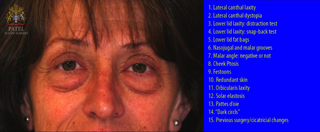
- Lower lid position - The position of the lower lid in the primary gaze is assessed at the limbus. Slight variations depending on the size of the eyeball or race are seen. Any cause of lid retraction such as prior surgery, thyroid orbitopathy, cicatricial skin diseases like pemphigus, Steven-Johnson syndrome, chemical burn, etc., must be explored in great detail. Any preexisting lid asymmetry should be explicitly pointed out to the patient and documented in photographs.
- Lower lid margin - Look for any active blepharitis or meibomian gland dysfunction.
- Snap-back test - Pull the lid inferiorly. Normal lids snap back to the original position immediately; the longer it takes to snap back, the more laxity is present. This should be incorporated into the surgical plan.
- Distraction test - Distract the lower lid forward using thumb and index finger. Any distraction more than 2 mm from the globe is considered abnormal.
- Lateral canthus laxity - Pull the lower lid medially away from lateral canthus. Normal displacement can be 0 to 2 mm; the greater the displacement of lateral canthus more laxity of the canthal tendons is present.
- Preoperative grades of the lower lid (based on both snap-back and distraction test) before performing lower blepharoplasty who require ‘tightening/anchoring procedure’[10]
- Grade +1 (Mild): Snap-back test - 1-2 seconds; Distraction test - 2-4 mm
- Grade +2 (Moderate): Snap-back test - 2-4 seconds; Distraction test - 4-6 mm
- Grade +3 (Severe): Snap-back test - next blink; Distraction test - greater than 6 mm
- Medial canthal laxity, punctal version, or punctal patency should be checked.
- Lower lid fat pads medial, central, and lateral should be assessed. The presence of the tear trough and lid-cheek junction is noted.
- The relation of the center of the cornea to the most prominent part on the cheek should be assessed. Normally they lie at the same level. Any recession of the cheek or negative malar vector may cause lid retraction after any blepharoplasty.
- Skin changes like thinning, the position of wrinkles, crow feet, pigmentation, festoons, etc., are also noted in eyelids and surrounding skin. Redundancy of skin assessed by a pinch test.
Preoperative Investigation
Systemic investigations can include the measurement of blood pressure, blood sugar levels, electrocardiogram, thyroid profile (if required). Patients on anticoagulant therapy should stop them for 1 to 2 weeks.
Patient Preparation
Local antiseptic solution (povidone-iodine ophthalmic paint) is used to clean the area to be operated. Skin markings are made using a marker pen and always in an upright, sitting position. The prominent fat pads and their margins should be marked along with the nasojugal and malar groove. Intravenous sedation is to be given (if planned, or general anesthesia, if planned). Local anesthesia (1% lidocaine with 1:100,000 epinephrine) is infiltrated in the marked area. An infraorbital nerve block can also be performed. The lateral orbital rim area can also be infiltrated if lateral canthal tightening procedure is to be performed, and topical ophthalmic tetracaine drops can be used to anesthetize the cornea and conjunctiva.
Technique or Treatment
Incision and Dissection
The skin is incised 1 to 2 mm below the lash line or within the prominent natural skin crease (if present) to hide the scar. Care should be taken to never extend the incision laterally beyond the orbital rim to prevent surgical stigmata. The incision should include only skin, not the orbicularis muscle. A Frost suture is placed at the grey line to protect the globe and facilitate retraction. Placing a corneal shield is optional and at the discretion of the operating surgeon.
Based on the anatomical correction planned, a skin-only or skin-muscle flap is elevated using blunt and sharp dissection. Hemostasis at every step is of the utmost importance using bipolar electrocautery under low power. If a skin-only flap is planned, the dissection below the skin is carried out to the inferior orbital rim or the last wrinkle. A skin-muscle flap can be elevated via the same subciliary skin incision with the following adjustments: the dissection below the skin is carried out to 4 to 5 mm below the lash line, preserving pretarsal orbicularis. Then orbicularis is divided, and an avascular plane below the preseptal orbicularis and above septum is reached, and dissection is carried to the inferior orbital rim. Great care should be taken not to breach the septum while dissecting.
Various modifications of traditional transcutaneous blepharoplasty have been described in the literature. McCollough places the skin incision inferior to the tarsal margin, thus preserving a cuff of pretarsal orbicularis oculi muscle, which plays an important role in blinking and tear drainage.[11] Another interesting modification includes a ‘fat preserving’ technique in which the herniated fat is pushed back into orbit, and capsulopalpebral fascia is sutured to the lower orbital rim, which strengthens it like a hernia repair.[12] A prospective comparative study comparing the ‘fat preserving’ approach and ‘traditional approach’ of 26 patients showed comparable aesthetic outcomes but lower recurrence and absence of the typical hollowing of the lower lid or sunken appearance of the globe in the ‘fat preserving’ group.[13][14]
Orbital Fat
The marking of fat pads should be done preoperatively and always in an upright, sitting position to determine the precise volume and location of fat pads to be excised and to determine whether post-septal or retro-septal fat needs to be preserved or transposed. This is of particular importance since over-resection of post-septal fat can lead to negative effects like a hollowed or cadaveric look.
If skin-flap is elevated, a button-hole incision of the orbital septum and orbicularis is made to reach medial, central, and lateral fat pads separately. If a skin-muscle flap technique is employed, all the fat compartments can be exposed at once through a single incision in the septum. Excess orbital fat easily pops out and can be teased using forceps and gentle, blunt dissection. Excess fat can be cross-clamped with a hemostat and then resected using insulated fine-tipped electrocautery. Complete hemostasis before leaving the resected fat stalk is mandatory. Care should be taken while resecting medial and central fat pads as inferior oblique muscle lies between them and can potentially be injured.
The arcus marginalis and the attachments of the orbicularis muscle can be released from the medial and central aspect of the inferior orbital rim to blunt the tear trough. After the release of arcus marginalis, supra-periosteal dissection is carried out to create pockets. The medial and central post-septal fat can be transposed over the orbital rim into the pockets and secured with 6-0 polyglactin sutures to fill the tear-trough further.[15] Hamara also described a ‘septal reset’ technique wherein, after the release of the septum from arcus marginalis, the septum is resutured to the inferior orbital rim, smoothing the lid-cheek junction.[16]
A modified transcutaneous lower lid blepharoplasty technique described by Huang uses a subciliary elliptical skin excision, and the medial, central, and lateral orbital fat pads are resected and then replanted via a micro-autologous fat transplantation technique to recontour prominent nasojugal and malar grooves, giving a rejuvenated appearance.[17]
Another alternative technique described by Liapakis et al. includes a subciliary approach for fat transposition and mid-face lift. In this technique, the nasal fat pad is repositioned with suturing at the medial canthus along with a lateral canthopexy to stabilize the lower lid. then a cheek flap is created and passed through a tunnel, and suspended to the periosteum of the lateral orbital rim.[18]
A percutaneous lower blepharoplasty technique has recently been described by Guner, consisting of removal and transfer of lower orbital fat pads through 4-mm skin incisions with minimal dissection.[19]
Orbicularis Oculi Muscle
Following the management of orbital fat, excess orbicularis muscle is elevated and resected, giving smooth contour. Hamara described a technique that utilizes the orbicularis muscle to support the lower lid. A laterally based flap of redundant orbicularis is suspended to the lateral orbital rim via upper lid incision.[15] A similar technique described by McCord[20] includes the fixation of pretarsal orbicularis along with skin to the inner aspect of the lateral rim.
Lateral Canthal Support
Lower lid laxity, if left undiagnosed and untreated while performing lower lid blepharoplasty, can lead to potential complications, including ectropion and lower lid retraction. The snap-back test and lid distraction test should be performed preoperatively to identify candidates for lateral canthal tightening. Various suture canthopexy and canthoplasties described in the literature can be chosen based on the patient’s need and the surgeon’s preference.[21][22][23][20]
Skin
Excess lower lid skin excision should be performed cautiously, as overzealous excision can lead to lid retraction and scleral show. The lower lid skin is gently placed over the lower lid structures, and skin is held vertically at the level of lateral canthus without any tension; this point is fixed with a suture. The excess skin medial and lateral to this point of fixation at lateral canthus is excised at the level of subciliary incision. The skin edges are sutured with 6-0 polypropylene or 6-0 polyglactin. In the case of a skin-muscle flap, the excess pre-septal orbicularis is excised and approximated with pretarsal muscle followed by skin excision and suturing.
Some surgeons prefer using ‘pinch blepharoplasty’ using transcutaneous subciliary incision to remove a pinch of excess skin with transconjunctival removal of fat, avoiding incision in the middle lamella.[10]
Complications
Retrobulbar Hemorrhage
One of the most feared complications in cosmetic surgery is vision loss, which in blepharoplasty is most likely to be due to retrobulbar hemorrhage causing optic nerve/retinal ischemia secondary to raised intra-orbital pressure. Though retrobulbar hemorrhage is well-described, vision loss is thankfully very rare. The majority of these cases present within the first 24 hours, especially in the first 3 hours after surgery.[24] The reported incidence of orbital hemorrhage after cosmetic peri-ocular surgery to be 1:2,000 and incidence of vision loss after hemorrhage 1:22,000 in this series. Symptoms include periorbital edema, pain, and vision loss. The pain is usually severe and may be associated with diplopia and vomiting. Signs include tense proptosis, chemosis, raised intraocular pressure, and loss of the pupillary light reflex. Any patient complaint of acute or severe eye pain in the immediate postoperative period after blepharoplasty warrants emergent examination, and a retrobulbar hemorrhage must be assumed until proven otherwise. Progressive retrobulbar hemorrhage may cause raised intraocular and intraorbital pressure involving superior orbital fissure and orbital apex, causing vision loss. Early recognition and prompt treatment are the keys to saving the eye. The aim of treatment is to reduce intraorbital and intraocular pressure. Surgical treatment includes emergent opening and exploring the wound for hematoma evacuation and cauterizing the offending vessel. Opening the surgical wound is also the best approach to decompress the orbital pressure. Lateral canthotomy and inferior cantholysis may be required to relieve the pressure in the orbit. Medical treatment alone is not indicated if a retrobulbar hematoma is present, as it will be far too slow to take effect.
Infection
It is a rare complication, though patients with diabetes and immunocompromised patients are at increased risk. Early infections can present with edema, erythema, pain, and fever. It may progress to cellulitis and abscess formation. The patient is managed on intravenous broad-spectrum antibiotics.
Diplopia
It can be transient or permanent. Transient edema can cause diplopia, which will resolve within a few days. Permanent diplopia is a result of damage to the inferior oblique muscle and will require surgical correction. This is thankfully very rare.
Lower Lid Malposition
Complications such as scleral show, lid retraction, and ectropion are possible with a subciliary approach. Milder grades of scleral show can be treated conservatively with massage and steri-strips. Ectropion in the early postoperative period is most likely due to unaddressed lower lid laxity, which should be identified preoperatively and corrected with lid tightening procedures. Ectropion can also occur in a more delayed fashion after over-resection of skin, which may sometimes require skin grafting. It is prudent to be very cautious in resecting skin in the lower eyelids.
Corneal Abrasion
Ocular injury includes corneal abrasion, ulceration, etc., which are rare. Topical antibiotic drops with patching may be required.
Dry Eyes
Temporary dryness is common, which usually will resolve 1 to 3 weeks after surgery. Topical lubricants are required in symptomatic patients.
Clinical Significance
Blepharoplasty surgery forms the cornerstone in the rejuvenation of the periorbita and, ultimately, the overall face. Transcutaneous lower lid blepharoplasty is a time tested, reliable and efficient procedure for correcting excess lower lid skin and fat bags.
Enhancing Healthcare Team Outcomes
The lower lid blepharoplasty is challenging as compared to upper lid blepharoplasty. Meticulous patient selection, preoperative evaluation, and surgical planning are important. The blepharoplasties are performed by multiple specialties, including ophthalmologists, oculoplastic surgeons, facial plastic surgeons, plastic surgeons, oral and maxillofacial surgeons, and otorhinolaryngologists. A thorough anatomical and physiological understanding of eyelid structure are necessary before attempting lower blepharoplasty. The interprofessional team for blepharoplasty is comprised of the operating surgeon, anesthesiologist, surgical assistants, and nursing staff. A well-coordinated team will lead to the best surgical outcome. Managing intraoperative and postoperative complication majorly depend on all team members. Postoperative wound care by trained nursing staff and proper counseling by surgeons have key importance for patient management.[5] [Level 5]
Nursing, Allied Health, and Interprofessional Team Interventions
The postoperative care by nursing staff includes cold compresses for 24-48 hrs to control edema. The patient should be closely observed for any deterioration in vision in the first 24 hours. Postoperative instructions include head elevation and avoidance of strenuous activity to decrease edema, lower intraocular pressure, and to decrease chances of hemorrhage. Oral analgesics and antibiotics are prescribed in the early postoperative period. Ocular lubrication is recommended in all patients and is particularly important in patients with preexisting dry eyes or post-surgery lagophthalmos. Along with lubrication, topical antibiotic-steroid ointment can be given to prevent and resolve chemosis.
Nursing, Allied Health, and Interprofessional Team Monitoring
The monitoring of any visual deterioration, periorbital edema, and ecchymosis is of utmost importance to recognize progressive retrobulbar hemorrhage in the early postoperative period. Close follow up, and proper wound care is essential by the surgeon and nursing staff.