Continuing Education Activity
A Galeazzi fracture is a fracture of the middle to distal third of the radius associated with dislocation or subluxation of the distal radioulnar joint (DRUJ). Advances in radiography and fracture research have helped define, classify, and guide operative management. Galeazzi fractures remain difficult to diagnose clinically, and debilitating complications can occur if proper treatment is not implemented. This activity reviews the evaluation and management of Galeazzi fractures and highlights the role of the interprofessional team in caring for affected patients.
Objectives:
- Identify common physical exam findings of a Galeazzi fracture.
- Describe the typical imaging findings associated with a Galeazzi fracture.
- Explain the treatment and management options available for Galeazzi fracture.
- Summarize interprofessional team strategies to improve care coordination and communication to advance the diagnosis and management of Galleazzi fractures.
Introduction
The forearm is an essential structure in the human body crucial for completing activities of daily living. It is designed to help maximize versatility by allowing pronation and supination of the hand. Forearm fractures can lead to significant short-term and long-term disability, mainly if treated incorrectly. Sir Astley Cooper first described this particular fracture in 1822, but it was not until 1934 that the eponym took hold when Riccardo Galeazzi presented the mechanism, incidence, and management of this injury. The Galeazzi fracture is a fracture of the middle to distal one-third of the radius associated with dislocation or subluxation of the distal radioulnar joint (DRUJ). Advances in radiography and fracture research have helped define, classify, and guide operative management. Galeazzi fractures remain difficult to diagnose clinically, and debilitating complications can occur if proper treatment is not started.[1][2][3]
Etiology
Galeazzi fractures most commonly result from a fall onto an outstretched hand with an extended wrist and hyperpronated forearm. The energy from the radius fracture gets transmitted towards the radioulnar joint leading to dislocation of the DRUJ. These fractures occur with a bimodal distribution, diaphyseal forearm fractures in young males are commonly due to high-energy trauma (e.g., sports injuries, falls from height, motor vehicle collisions) and fractures in aging females are due to low-energy traumas such as falls from ground level.[4][5][6]
Epidemiology
Galeazzi fractures account for approximately 7% of all forearm fractures in adults. One in four radial shaft fractures is a true Galeazzi injuries. Distal forearm fractures are far more common than midshaft forearm fractures, which occur in about 1 to 10 per 10,000 people per year. The peak incidence in children is nine to 12 years. The most noteworthy risk factors for midshaft forearm fractures include sports (football and wrestling), osteoporosis, and being post-menopausal. These risk factors correlate with a bimodal incidence with the highest occurrence in young males (10:10,000) and elderly females (5:10,000).
Pathophysiology
Anatomy
The osseous forearm is composed of the radius and ulna bones. The radius and ulna are stabilized by three groups of ligamentous structures: distally the triangular fibrocartilage complex (TFCC), the interosseous membrane, and proximally the annular ligament. The interosseous membrane is responsible for dispersing axial load force to the forearm, 60% to the radiocapitellar joint and 40% to the ulnohumeral joint. The radiocapitellar joint largely stabilizes the proximal forearm while the TFCC predominantly supports the distal forearm. Proximally the ulna consists of the olecranon and coronoid. Distally the ulnar head serves as an insertion point for the TFCC, supplementing the DRUJ. The proximal radial head articulates with the capitellum of the humerus (radiocapitellar joint), rotating within the annular ligament during pronation and supination. Distally the radius connects with the lunate and scaphoid bones of the wrist.
Classification System
Two classification systems have been proposed when categorizing Galeazzi fractures.
The first classifications were based on the position of the distal radius:
Type I: Dorsal displacement
Type II: Volar displacement
The second classification system is based on Rettig ME and Raskin KB who categorized Galeazzi fractures based on fracture stability. They found stability to be dependent on the distance of the radial fracture from the distal radial articular surface:
Type I:
- Fracture occurring distally from the 7.5 cm demarcation (i.e., closer to the wrist)
- Associated with significant DRUJ instability in more than 50% of cases
Type II:
- Fracture occurring proximally from the 7.5 cm demarcation (i.e., further from the wrist)
- Associated with significant DRUJ instability in only around 5% of cases
History and Physical
Patients with diaphyseal forearm fractures typically complain of pain at the site of injury. An examination should begin with a visual inspection of the skin and soft tissue paying close attention to visible bony deformities, skin lacerations, muscle contusions, tendon damage and neurovascular deficits. It is essential to identify wounds overlying fracture sites (i.e., open fracture), which necessitates immediate surgical intervention. Gentle palpation should be performed to identify deformities and focal tenderness. Stability of the proximal and distal joints should be assessed to identify concomitant injuries. A fall on an outstretched hand should raise suspicion for a wrist injury, and particular attention should be paid to the stability of the DRUJ. Avoid probing open wounds. High mechanism crush injuries merit a detailed neurovascular exam with repeat serial exams looking for signs of acute compartment syndrome. Inquire about weakness, numbness, paresthesias, and radiating pain. Although nerve injury is less common, examination of the median and radial nerve distribution is essential in identifying nerve damage. Ulnar nerve injury is rare.
Evaluation
Imaging
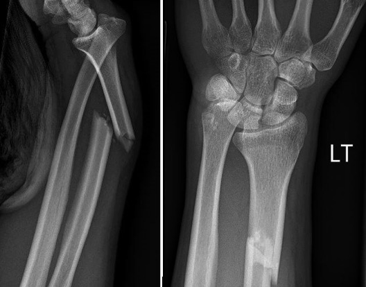
If a forearm fracture and dislocation are suspected, radiographs are warranted. An anteroposterior and lateral view will usually identify the injury. An additional oblique view may help better classify the injury. Additional radiographs of the distal wrist and proximal elbow should be obtained with any suspicion of coexistent injury.
If a distal to mid-shaft radial fracture is seen on the radiograph, close examination of the DRUJ is merited. Signs of DRUJ disruption include:
- Widening of the DRUJ on the PA view
- Ulnar styloid fracture
- Displacement of the ulna dorsally on the lateral view
- Radial shortening greater than 5 mm (would need to compare with unaffected limb)
Usually, advanced imaging is not needed for initial assessment. For pre-operative planning, a CT scan may be used to evaluate for non-union, and magnetic resonance imaging (MRI) can help evaluate for TFCC tears and interosseous membrane disruption.
Treatment / Management
All suspected or confirmed Galeazzi fractures will require orthopedist consultation. While awaiting consult, patients should be placed in a sugar-tong splint. In most cases, conservative management is indicated in children while surgical intervention is warranted in adults. Prior reports suggested treatment with closed reduction and immobilization alone, in adults, yielded poor outcomes in greater than 90% of patients.[7][8]
Initial Management
Initial management for a presumed fracture includes rest, ice, immobilization, and elevation. In most cases, closed reduction of the radius followed by reduction of the ulna in the DRUJ should be attempted in the acute setting.
Pediatrics
Children tend to have overall better long-term outcomes compared to adults. The approach is usually conservative with closed reduction and splinting. Above-elbow casting in supination is the preferred immobilization. Irreducible and unstable injuries, as well as variants of the Galeazzi fracture, may require surgical intervention with open reduction and internal fixation (ORIF).
Adults
Adults tend to have poor outcomes with closed reduction and immobilization. Multiple reports have shown high rates of nonunion and secondary displacements with conservative management. Surgical intervention is the preferred intervention. The radial shaft fracture is first repaired with rigid fixation. Intra-operatively, after rigid fixation of the radius, the DRUJ stability should be assessed while in supination. If the joint is stable, two K-wires pinned. If instability is noted, the TFCC should be repaired followed by pinning. If an ulnar styloid fracture accompanies the unstable DRUJ, the TFCC should be repaired followed by fixation of the styloid using a lag screw or tension band wire. Patients with irreducible DRUJ will require open reduction with the removal of obstructing soft tissue and repair of the TFCC. Patients are placed in an above the elbow splint/cast in supination.
As with all fractures, length of recovery is dependent on multiple variables including the severity of the injury, individual's ability to heal, and the intended use of the extremity. Rehabilitation usually begins six to eight weeks after surgical fixation. The goal of rehab is the return of full range of motion and fine motor skills with the absence of pain. Full return to activity depends upon the severity of the injury as well as the patient’s intended use of their upper extremities. Manual workers and athletes may require more prolonged rehabilitation. Typical return to full activity in patients with low physical demands can happen after 8 to 12 weeks. Patients with high demand activity (manual workers and athletes) may need up to 12 to 16 weeks of rehab. Surgical hardware is usually permanent, with less than 10% of patients needing hardware removal.
Differential Diagnosis
- Elbow dislocation in emergency medicine
- Emergent management of hand dislocation
- Hand fracture management in ED
- Wrist dislocation in emergency medicine
- Wrist fracture in emergency medicine
Pearls and Other Issues
Galeazzi injuries are associated with very high morbidity and are best managed by a multidisciplinary team that includes an orthopedic surgeon, emergency department physician, therapist, orthopedic nurse and the radiologist. Galeazzi fractures are easily missed, particularly the DRUJ dislocation, as they can be difficult to identify on radiographs. When patients present with forearm and elbow fractures, an orthopedic consult should be made.[9][10]
The complexity of Galeazzi fractures leads to a variety of outcomes. It is estimated that the overall complication rates of Galeazzi fractures are close to 40%. Children usually fare better than their adult counterparts. As time passes without the correct identified diagnosis, the more difficult it becomes to manage the injury, and the patient is more likely to have significant morbidity.
Forearm midshaft fractures have malunion or nonunion occurring in approximately 2% to 10% of cases, which is higher than the average forearm fracture nonunion rate (2%). Other notable complications include acute compartment syndrome (pre- and post-surgical), radioulnar synostosis (1% to 6%), elbow stiffness from protracted immobilization (adults), myositis ossificans, ulnohumeral osteoarthritis, and wound infections (0% to 3%).
Muscle-tendon entrapment may occur with significant injuries. The extensor carpi ulnaris, extensor digiti minimi, and flexor pollicis longus tendons are the most common to become entrapped with Galeazzi fractures. They can make reduction difficult and may require surgical repair to avoid chronic instability.
Branches of the radial nerve are the most common neurovascular structures to get injured. This is usually due to laceration of the radial sensory nerves during a volar surgical approach. The most common associated motor deficit is from injury to the posterior interosseous nerve, which can get damaged during a dorsal surgical approach. Transient injury to the anterior osseous nerve branch of the median nerve has been reported. Ulnar nerve injuries are rare. Nerve injuries rarely require treatment, and the majority of patients have complete resolution of symptoms in 9 to 12 weeks.
Enhancing Healthcare Team Outcomes
Galeazzi injuries are associated with very high morbidity and are best managed by an interprofessional team that includes an orthopedic surgeon, emergency department physician, therapist, orthopedic nurse and the radiologist. Galeazzi fractures are easily missed, particularly the DRUJ dislocation, as they can be difficult to identify on radiographs. When patients present with forearm and elbow fractures, an orthopedic consult should be made.
The complexity of Galeazzi fractures leads to a variety of outcomes. It is estimated that the overall complication rates of Galeazzi fractures are close to 40%. Children usually fare better than their adult counterparts. As time passes without the correct identified diagnosis, the more difficult it becomes to manage the injury, and the patient is more likely to have significant morbidity.
Even after treatment, recovery is often prolonged and rehabilitation is required to regain muscle strength and function.[3][11]