Continuing Education Activity
Rectal prolapse refers specifically to the prolapse of some or all of the rectal mucosa through the external anal sphincter. In pediatric populations between infancy and age 4, rectal prolapse is usually a self-limiting condition, responding to conservative management. The highest incidence of rectal prolapse has been noted in the first year of life. However, children presenting after age 4 usually have a chronic condition predisposing them to have developed rectal prolapse. This activity reviews the cause, pathophysiology, and presentation of rectal prolapse and highlights the role of the interprofessional team in its management.
Objectives:
Review the etiology of rectal prolapse.
Describe the evaluation of a patient with rectal prolapse.
Summarize the treatment options for rectal prolapse.
Explain the importance of improving care coordination among interprofessional team members to improve outcomes for patients affected by rectal prolapse.
Introduction
Rectal prolapse refers specifically to the prolapse of some or all of the rectal mucosa through the external anal sphincter. In pediatric populations between infancy and age 4, rectal prolapse is usually a self-limiting condition, responding to conservative management. The highest incidence of rectal prolapse has been noted in the first year of life.[1] However, children presenting after age 4 usually have a chronic condition predisposing them to have developed rectal prolapse.[2] In some cases, prolapse may persist indefinitely, requiring surgical intervention.[3]
Etiology
Pediatric populations are more likely to develop rectal prolapse due to several anatomic differences in early childhood. In children, the rectum follows a vertical course along the sacrum and coccyx and is also in a relatively lower position than the other pelvic organs. The rectum also has a rather redundant rectal mucosa, which is loosely attached to the underlying muscularis. The sigmoid colon has more mobility than adult populations, and the levator ani muscle offers relatively little support. Finally, Houston valves, which provide structural integrity in the rectum, have not yet fully developed in the majority of infants less than one year.[1]
Rectal prolapse is also caused by increased bowel motility, increased abdominal pressure, and certain congenital conditions, to be outlined below. Increased bowel motility occurs most secondarily to infectious diarrhea caused by amebiasis, giardiasis, trichuriasis, Salmonella, Shigella, and Escherichia coli 0157:H7. Ulcerative colitis and laxative abuse may also cause increases in bowel motility. Next, increased abdominal pressure may commonly be caused by chronic constipation, protracted coughing, excessive vomiting, or straining at urination because of outlet obstruction. Finally, congenital conditions such as cystic fibrosis, myelomeningocele, Hirschsprung disease, spina bifida, and congenital hypothyroidism are more likely to develop rectal prolapse. Other causes include malnutrition and anatomic defects such as mucosal polyps or tumors and imperforate anus post-repair.[1][4]
Epidemiology
In pediatric populations, rectal prolapse occurs equally in male and female patients.[1] The highest incidence occurs from ages 1 to 3.[5] The affliction is much more common in underdeveloped countries, with common causes including parasitic disease, malnutrition, and diarrheal illness. In the United States, constipation is the most common association.[1]
Pathophysiology
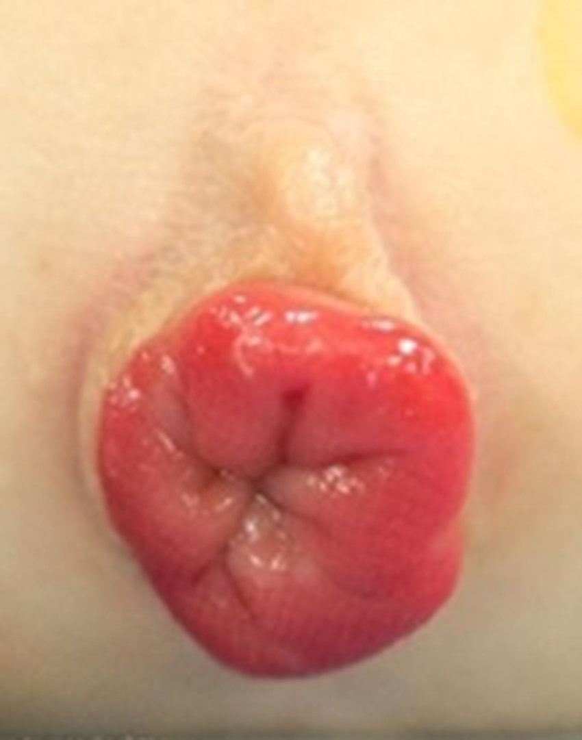
As noted above, rectal prolapse refers specifically to the prolapse of some or all of the rectal mucosa through the external anal sphincter. There are 2 types of rectal prolapse: type 1 and type 2, also called false procidentia and true procidentia, respectively. Type 1, which is partial or mucosal prolapse, produces radial folds at the junction with the anal skin. This type usually involves less than 2 cm of prolapse, and only the mucosa is prolapsed. Type 2, or complete prolapse, is characterized by full-thickness extrusion of the rectal wall. Concentric folds are seen in the prolapse mucosa. This type of prolapse, which is similar to intussusception functionally, is further divided into first, second, and third-degree prolapse. First-degree type 2 prolapse protrudes greater than 5 cm from the anal verge and includes the mucocutaneous junction. Second-degree prolapse protrudes only 2 to 5 cm from the anal verge. Finally, third-degree prolapse or occult rectal prolapse is an internal process and thus does not protrude through the anal verge.[1][4]
History and Physical
In adolescents with rectal prolapse, symptoms include tenesmus, anorectal pain, and passage of blood and mucus. In children, rectal prolapse is typically brought to medical attention after being detected by the patient’s parents. A dark red mass with or without mucus and blood that protrudes from the rectum during straining is described, yet this is commonly spontaneously resolved by the time the patient presents. Prolapse is usually painless or associated with only mild discomfort. At the time of prolapse, decreased or absent rectal tone may be present on a digital rectal exam, but the tone is usually normal after a few hours.[1]
Evaluation
Diagnosis of rectal prolapse is most commonly made based on history and physical alone. As mentioned above, prolapse often resolves by the time the patient reaches medical attention, and thus, the clinician must rely on history for diagnosis. Patients with constipation as the likely cause of their prolapse should receive contrast radiography of the colon and anorectal manometry.[6] When evaluating third-degree or occult rectal prolapse, colonoscopy or sigmoidoscopy often reveals erythematous granularity of the distal rectum, in addition to a polypoid white-topped mucosal lesion on the anterior rectal wall.[7] These tests may also help visualize rectal polyps or ulcers if present. Evaluation for associated pelvic floor anomalies and further characterization of prolapse may also per performed by fluoroscopic dynamic defecography or magnetic resonance imaging.[1]
Treatment / Management
Conservative management of rectal prolapse includes stool softeners and/or laxatives, avoidance of prolonged straining, and treatment of any predisposing underlying conditions. Compliance with bowel regimens is important because patients who have repeat instances of rectal prolapse may be less responsive to rectal prolapse in the future. Generally, these conservative tools work for about 90% of patients who develop rectal prolapse before age 3. For children with cystic fibrosis, who, as mentioned above, are predisposed to rectal prolapse, adjustment of pancreatic enzymes is important.[1] Further management is indicated when patients complain of longstanding symptoms, rectal pain, rectal bleeding, rectal ulceration, or difficult manual reduction of the prolapsed rectum.[8] Injection sclerotherapy is often the first intervention, followed by Thiersch cerclage and rectopexy.[1]
Differential Diagnosis
The differential diagnosis for rectal prolapse includes ileocecal intussusception, prolapsing rectal polyp, prolapsing rectal duplication cyst, and rectal hemorrhoids. While rectal prolapse is painless, intussusception presents with intermittent severe pain. Examination of prolapse tissue can distinguish between prolapsing rectal polyp, prolapsing rectal duplication cyst, and rectal hemorrhoids due to the circumferential nature of prolapse.[1]
Prognosis
The prognosis of rectal prolapse is generally good, especially when diagnosed between 9 months and 3 years, and not associated with any other underlying condition. It is usually a childhood condition and often does not recur after age 6. When rectal prolapse is diagnosed after age 4, children are more likely to have underlying neurologic or musculoskeletal problems requiring surgical management and symptoms into adulthood.[1][4]
Complications
Most complications are from surgery and may include:
- Bleeding
- Damage to the bladder neck
- Damage to the presacral nerves
- Pelvic hematoma
Enhancing Healthcare Team Outcomes
Rectal prolapse is ideally managed by an interprofessional team that involves a pediatrician, gastroenterologist, pediatric surgeon, and a pediatric nurse. All children with rectal prolapse should undergo testing for cystic fibrosis. While awaiting surgery, parents should be taught how to reduce rectal prolapse.[9][10]
Coordination of care for rectal prolapse requires a highly organized interprofessional team. For diagnosis and conservative management, physicians, nurses, and families must work together for effective treatment and maintenance at home. If these treatments are effectively communicated and orchestrated, the patient will ultimately have a better outcome. If surgery is indicated for prolapse, coordination is also needed between physicians, nurses, pharmacists, and other members of the healthcare team. The importance of communication, patient-centered care, and professionalism cannot be understated when working with pediatric populations.[11]