Introduction
The middle cranial fossa, also known as the central skull base, is an intricate intracranial area that contains many structures susceptible to pathology, making it an area directly pertinent to neurosurgeons, neurologists, otolaryngologists, ophthalmologists, radiologists, and endocrinologists.[1][2] The middle cranial fossa can be divided into medial and lateral portions, each containing many nerves, vessels, and cranial structures crucial to its function. In addition to its contents, the middle cranial fossa acts as a potential space for infection and hemorrhage. The complex anatomy of this region makes it a difficult area for surgeons to traverse, but also provides access to various areas of the brain for a variety of procedures. The following review article will highlight the complex structure of the middle cranial fossa while simplifying the contents and various pathologies that affect structures within the middle cranial fossa.
Structure and Function
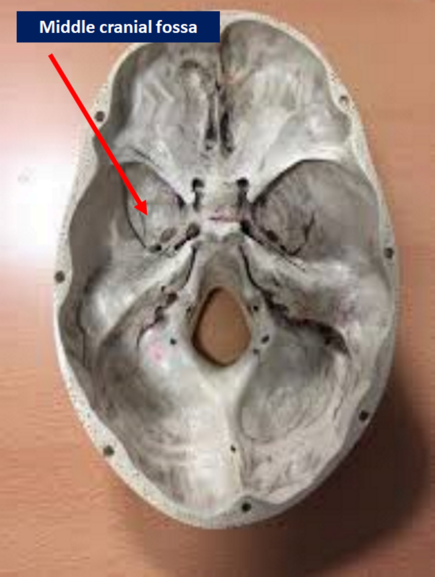
Generally, the middle cranial fossa is described as having a “butterfly” appearance when viewed from above. The middle cranial fossa is a paired structure that fuses medially to form the sella turcica. Each side of the middle cranial fossa consists of the sphenoid and one temporal bone.[3] The middle cranial fossa is bound anterolaterally by the lesser wings of the sphenoid and anteromedially by the limbus of the sphenoid.[4] Posterolaterally, it is bound by the petrous ridge and posteromedially by the dorsum sellae. It is also laterally bound by the greater wings of the sphenoid, squamous temporal bone, and sphenoidal angles of the parietal bones. With its complex structure, the middle cranial fossa functions to house a wide variety of nerves and vessels as they pass through the many foramina present in the fossa.[3][4] Between the two middle cranial fossa sides, a concave depression, known as the sella turcica, houses the pituitary gland. To each side of the sella turcica, the internal carotid artery (ICA) and multiple cranial nerves pass through the cavernous sinus. The cavernous sinus acts as a potential space for infection, hemorrhage, and thromboses that will receive attention in later sections.[5] The central aspects of the middle cranial fossa contain the horns of the temporal lobes along with many foramina that transmit multiple vessels and nerves. The lesser wings of the sphenoid span to the lateral edges of the middle cranial fossa and function as an attachment point of the tentorium cerebelli.[6]
The structure and function of the middle cranial fossa center around the foramina that transmit a variety of cranial nerves and vessels. In the anterior midline of the middle fossa within the sphenoid bone, the optic foramina open into the optic canals which ultimately terminate into the orbital cavities. Of clinical and surgical importance, the prechiasmatic sulcus is a depressed groove between the optic foramina and plays a role in skull base surgery due to its predisposition to hiding cranial base meningiomas. Due to this reason, extensive research has been conducted to establish the anatomic and radiological morphology of the sulcus.[4]
Within the medial aspect of the middle cranial fossa, from medial to lateral, there are four foramina: the superior orbital fissure, pterygoid canal, foramen rotundum, foramen ovale, and foramen spinosum. These foramina are in the sphenoid bone. The lateral portions of the middle cranial fossa form by the temporal bones and are defined by three important foramina: the carotid canal, hiatus of the greater petrosal nerve, and the hiatus of the lesser petrosal nerve. At the intersection of the sphenoid, temporal, and occipital bones, there is a foramen pierced by small vessels called the foramen lacerum.[7]
Embryology
While complex, the development of the skull base is divisible into a few basic principles. The middle cranial fossa, a portion of the skull base, is derived from neural crest and mesoderm by day 40 of gestation. The majority of the neural crest cells migrate from the first and second pharyngeal arches. These neural crest cells then condense and eventually undergo chondrogenesis and subsequent ossification. Interestingly, while most of the cranial vault ossify through intramembranous means, the skull base primarily undergoes endochondral ossification.[5] The skull base foramina form by cartilage fusing around pre-existing structures.[6]
Blood Supply and Lymphatics
An in-depth understanding of cerebral vascular anatomy is necessary for all clinicians. For example, one of the most crucial vessels for cerebral blood flow is the internal carotid artery, which has a complex course through the middle cranial fossa. The internal carotid artery enters the middle cranial fossa through the carotid canal, which lies posteromedial to the foramen ovale forming the petrous internal carotid artery as it courses along the petrous temporal bone. Eventually, it courses through the cavernous sinus where it runs medial to CN VI before it enters the circle of Willis in the cerebrum.[8]
The ophthalmic artery is a branch of the internal carotid artery which originates either inferior or inferomedial depending on the anatomical variation relative to the optic nerve, but after its exit, it travels lateral to the optic nerve as it traverses the optic canal. The ophthalmic artery’s anatomic relationship to the optic nerve is essential in various ophthalmologic and neurosurgical operations to avoid injury to the optic nerve and ophthalmic artery.[9]
The middle meningeal artery is a tributary of the maxillary artery and supplies the supratentorial dura mater along with the periosteum of the inner table of the cranial bones. This artery enters the middle cranial fossa through the foramen spinosum before taking a superficial course along the dura. The vessel superficially courses along the temporal bone, implicating it as a source of rapid arterial (epidural) bleeding in the setting of a traumatic temporal bone injury.[10]
Nerves
Many nerves traverse the middle cranial fossa through the foramina previously discussed. This section will discuss the intracranial anatomy of each of these nerves in cranial nerve numerical order. The first nerve passing through the middle cranial fossa is purely sensory: the optic nerve, cranial nerve (CN) II. Axons from the optic nerve originate from the ganglion cells of the inner retina, and these axons merge at the optic papilla where they form the optic nerve. There are four anatomic compartments of the optic nerve: intraocular, intraorbital, intracanalicular, and intracranial portions. In this activity, the focus will be on the intracranial portion. The intracranial portion begins when the nerve enters the optic canal at the terminus of the orbital apex. As the nerve passes through the optic canal, it takes an upward course at an approximately 45-degree angle, continuing until it forms the optic chiasm above the diaphragm sella and pituitary.[11]
The oculomotor nerve, CN III, is purely a motor nerve that triggers movements of the superior rectus, medial rectus, inferior rectus, inferior oblique, and levator palpebrae muscles. It also controls visceral efferent fibers, such as the sphincter pupillae and ciliary muscles.[12] The cavernous section of the oculomotor nerve is attached to the lateral wall of the cavernous sinus between the two dural “leaves” of the lateral sinus wall.[2] This anatomy is similar to the fourth, ophthalmic (V1), and maxillary (V2) cranial nerves, but they are different from the sixth cranial nerve, which will be discussed in a later section. The third and fourth cranial nerves travel in close proximity as they traverse the roof of the cavernous sinus en route to the superior orbital fissure. CN III runs superior to the internal carotid artery within the cavernous sinus and predisposes the nerve to compression by large cavernous internal carotid artery aneurysms; however, compression of CN VI is more common. [2] As the nerve exits the cavernous sinus, the fissural segment of the nerve begins as the nerve enters the medial superior orbital fissure. Approximately 2 to 3 mm before it enters the superior orbital fissure, it splits into superior and inferior divisions as the branches progress to their target muscles.[13][12]
The trochlear nerve, CN IV, is a motor nerve that triggers the movement of one extraocular muscle, the superior oblique. Anatomically, the nerve enters the cavernous sinus along the lateral sinus wall slightly inferior to the oculomotor nerve and superior to the ophthalmic division of the trigeminal and then passes through the superior orbital fissure to enter the orbit.[13]
The trigeminal nerve, CN V, is a mixed motor and sensory nerve that has a significant relationship to the middle cranial fossa as its ganglion and branches course through many middle cranial fossa structures. Of structural importance is the location of the trigeminal ganglion in the intracranial cavity within Meckel’s cave. Meckel’s cave is a mouth shaped depression in the middle cranial fossa, lateral to the cavernous sinus, that is continuous with the posterior cranial fossa and contains the trigeminal nerve, trigeminal ganglion, along with V1, V2, and V3 (mandibular) branches.[14] Stemming from Meckel’s cave, the V1 and V2 branches enter the cavernous sinus along the lateral sinus wall slightly inferior to the trochlear nerve. The branches of the trigeminal nerve then exit the middle cranial fossa through the superior orbital fissure (V1), foramen rotundum (V2), and foramen ovale (V3).[15]
The abducens nerve, CN VI, is simply a motor nerve that triggers the movement of one extraocular muscle, the lateral rectus, and has a unique anatomic course within the cavernous sinus. While the other cranial nerves that course through the cavernous sinus travel between the two dural “leaves” within the lateral walls of the sinus, CN VI travels unprotected within the sinus itself close to the inferolateral aspect of the internal carotid artery. Once through the cavernous sinus, the nerve then exits the middle cranial fossa through the superior orbital fissure.[13] The close proximity of CN VI to the cavernous internal carotid artery makes it more susceptible to injury and compression from the internal carotid artery or pathology within the cavernous sinus compared to other cavernous sinus structures.
There are two other smaller nerves that pass through the middle cranial fossa: the greater superficial petrosal nerve and the lesser superficial petrosal nerve. The greater superficial petrosal courses into the foramen lacerum and joins the deep petrosal nerve to form the vidian (pterygoid) nerve. There is surgical importance to this anatomical relationship since it can be traced posteriorly to the internal carotid artery and is a surgical landmark during skull base surgery. The lesser superficial nerve runs anterior and parallel to the greater superficial petrosal nerve, eventually exiting through the foramen petrosum, between the foramen spinosum and foramen ovale.[16][6]
Physiologic Variants
As physiological variants, we can find foramina of different shapes or with the presence of tubercles and bony plates, or in some cases, an absence of foramina, duplications and the presence of bone spines. [17]
Surgical Considerations
A variety of conditions affect the middle cranial fossa and an understanding of this area of the skull base is paramount for any clinician or surgeon encountering these conditions. A non-exhaustive list of conditions affecting the middle cranial fossa includes pituitary adenomas, vestibular schwannomas, cavernous sinus thrombosis, cavernous sinus infection, cranial nerve palsies, and intracranial meningiomas. The surgical approach is highly dependent on the location of the pathology addressed. Vestibular schwannomas involve the posterior cranial fossa; however, often a CN VIII sparing middle cranial fossa approach will be used to preserve hearing. In the case of a tuberculum sellae meningioma, surgical approaches ranged from bilateral subfrontal to endoscopic endonasal approaches with the course of surgery depending on the location, size, and surgeon’s preference. Recent literature has suggested that in the case of skull base meningiomas, combination surgical excision and stereotactic radiosurgery was acceptable management.[18][19]
Clinical Significance
It is impossible to overstate the clinical significance of the middle cranial fossa due to its contents and the numerous pathologies associated with these structures. In this section, some of the most common pathologies will get brief coverage. Cavernous sinus syndrome is characterized by ophthalmoplegia and cranial sensory deficits due to the involvement of multiple cranial nerves in the cavernous sinus. The pathologies that can cause cavernous sinus syndrome vary widely, but can include malignancy, infectious, inflammatory, granulomatous, or vascular etiologies. Regarding malignancy, head and neck tumors are the most likely to metastasize to the cavernous sinus. However, breast, lung, and prostate cancers have also been reported to metastasize to this area.[20]
Since the pituitary gland lies within the middle cranial fossa, many endocrine pathologies have associations with the middle cranial fossa. In the case of a pituitary microadenoma, excess hormone production is common, causing systemic symptoms associated with hormone hypersecretion. In the case of a pituitary macroadenoma, a majority of the symptoms have correlations with compression of nearby structures such as the optic chiasm, leading to bitemporal hemianopsia.[21]