Continuing Education Activity
Cor triatriatum is a rare congenital heart defect in which a thin, fibromuscular membrane causes division of either the right or left atrium, resulting in the presence of three atrial compartments. Infants are usually diagnosed during the first year of life, however, in some cases this disease is not diagnosed until adulthood. Patients may present with cough, dyspnea, wheezing, pulmonary congestion, tachycardia, heart murmur, palpitations, pallor, and/ or failure to thrive. This condition requires surgical management or heart failure may develop due to complications such as recurrent pneumonia and bronchial inflammation. This activity reviews the clinical evaluation of cor triatriatum and the role of the health professional team in coordinating the care of affected patients.
Objectives:
Explain when cor triatriatum should be considered on differential diagnosis.
Identify the best tests for cor triatriatum.
Review the management of cor triatriatum.
Describe how coordination of the interprofessional team can lead to more rapid diagnosis of cor triatriatum and subsequently decrease associated morbidity and mortality in affected patients.
Introduction
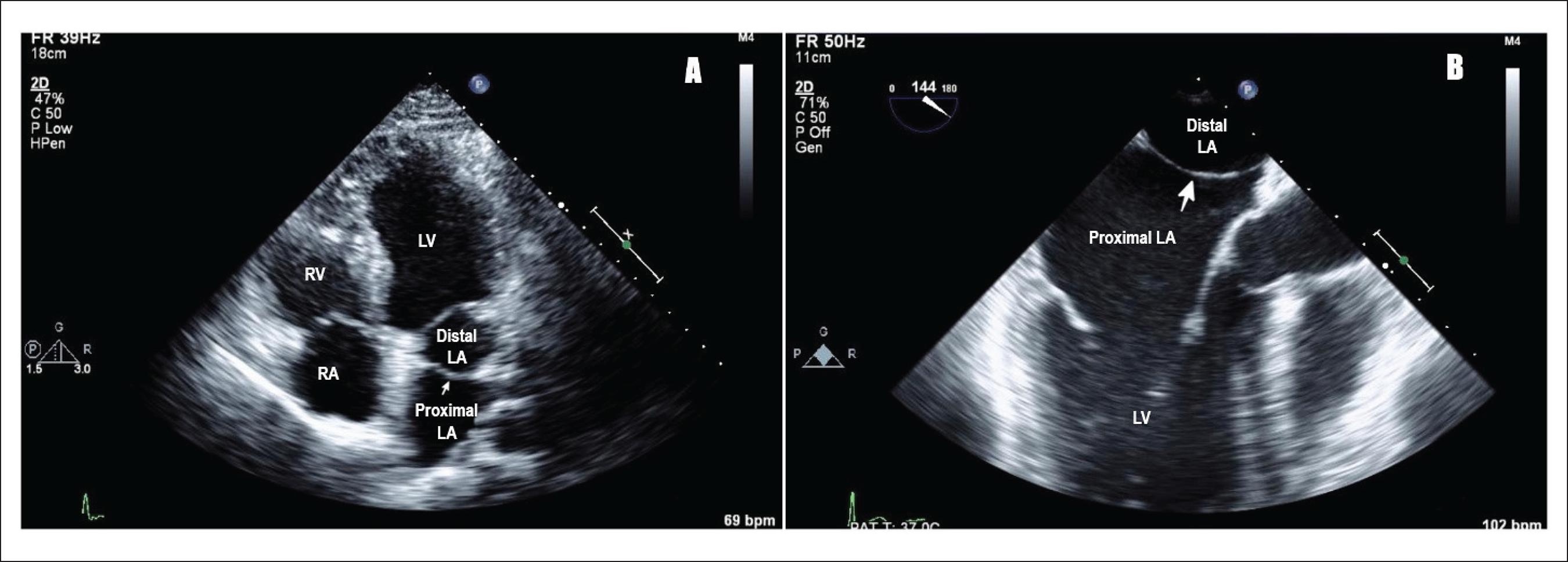
Cor triatriatum is a rare condition occurring when a child is born with a thin, fibro-muscular membrane subdividing either the left or the right atrium into 3 chambers (see Image. Cor Triatriatum). The condition is also classified as a congenital heart defect. Cor triatriatum sinister is the most common form. The left atrium divides via an atrial appendage into an upper and a lower chamber. The upper chamber receives blood from the pulmonary veins, while the lower chamber is attached to the left atrial appendage blocking the mitral valve orifice creating a significant left ventricular inflow obstruction. The presence of the left atrial appendage differentiates cor triatriatum from another congenital heart defect, supravalvular mitral stenosis.[1]
Another, rarer form of cor triatriatum is cor triatriatum dextrum. With cor triatriatum dextrum the right valve of the sinus venosus persists dividing right atrium into 2 chambers. This form presents similarly to Ebstein's anomaly and is difficult to differentiate.
Etiology
Cor triatriatum results from the incomplete absorption of the common pulmonary vein, which is normally reabsorbed during the development of a fetus and becomes a part of the left atrium. The incomplete absorption results in the formation of an appendage which subdivides the left atrium into 2 chambers. No known genetic mutations or risk factors are found to be associated with the development of this condition.
Epidemiology
Cor triatriatum presents in association with other congenital cardiac defects such as tetralogy of Fallot, atrial septal defect, ventricular septal defect partial anomalous pulmonary venous connection, and represents only 0.1% to 0.4% of all congenital abnormalities.[2] The membrane may be complete or may contain one or more fenestrations of differing size.
Pathophysiology
A malincorporation theory[3] presented by Dr. Clifford G. Parsons explains how cor triatriatum sinister occurs. During normal fetal development, the pulmonary vein incorporates into the left atrium. If it fails to do so, the common pulmonary venous ostium remains narrow, resulting in a septum-like structure called the atrial appendage. The appendage then further divides the left atrium into 2 compartments. Although widely accepted, this theory fails to explain how fossa ovalis and atrial muscle fibers are also present in the proximal atrial chamber.[4]
Two other theories, malseptation theory and entrapment theory, also explain the pathophysiology of cor triatriatum. The malseptation theory states that the fibro-muscular membrane is an abnormal growth of the septum primum; whereas, entrapment theory emphasizes the entrapment of the common pulmonary vein in the embryonic sinus venosus, thereby preventing its incorporation into the left atrium. The malincorporation theory remains the most widely accepted theory.
History and Physical
Classically, cor triatriatum presents in infancy with signs and symptom of pulmonary hypertension and pulmonary venous obstruction. Due to low cardiac output, children can show poor growth and weight gain, feeding difficulties, respiratory distress, and tet spells. In childhood and adulthood, the signs and symptoms of pulmonary venous hypertension and right heart failure dominate as the membrane calcifies, and the opening becomes smaller and smaller decreasing cardiac output even further. Mitral valve regurgitation and atrial fibrillation impose serious dangers. Most patients present with the following features[5]:
- Dyspnea and orthopnea
- Easy fatigability
- Hemoptysis
- Exercise intolerance and shortness of breath
- Palpitations
Atrial fibrillation can cause systemic thromboembolism and present as pulmonary embolism or stroke. The left atrial enlarges due to backing up of blood and can present as life-threatening arrhythmias.
Cor triatriatum sinister that presents for the first time in adulthood is rare but possible. It presents similarly to mitral stenosis, but the absence of loud S1 and an opening snap helps to distinguish between the 2. Chest x-ray showing pulmonary congestion with an absence of left atrial enlargement is characteristic of cor triatriatum. A continuous gradient on Doppler echocardiography confirms the diagnosis.[4][6]
Physical Exam
The physical exam findings are due to the right heart failure and pulmonary congestion.
- Accentuated pulmonary component of second heart sound
- A soft mid-systolic murmur at the upper left sternal border with a wide and fixed splitting of an atrial septal defect.
- Right ventricular heave
- Rales at the lung base
- Diminished peripheral pulses
- Hepatomegaly and right upper quadrant tenderness
- Ascites
- Peripheral edema
- Distended peripheral veins
- Distended jugular venous and elevated jugular venous pressure
- Pallor
- Poor weight gain
Evaluation
The mainstay of evaluation and diagnosis includes imaging studies such as chest x-ray, ECG and echocardiography, angiography and left and right heart catheterization.[7][8]
Chest X-Ray
A chest x-ray is the initial investigation of choice. The findings include:
- Pulmonary congestion with haziness (Kerley B-lines)
- Ground glass appearance of acute pulmonary edema
- Prominent pulmonary vessels
- Pleural effusion
- Left atrial enlargement
- Cardiomegaly
Electrocardiography (ECG)
ECG findings are non-specific in cor triatriatum. Can range from atrial fibrillation and no specific P-wave changes to right axis deviation due to pulmonary congestion and right ventricular hypertrophy.
Echocardiography
Echocardiography is the diagnostic modality of choice as it not only allows for definitive diagnosis, but the 3-dimensional reconstruction of echocardiographic images pinpoints the exact location of the defect and appendage, helps direct the surgical approach to the disease. The left atrial appendage and its fenestrations can be easily visualized along with the presence of an atrial septal defect, pulmonary stenosis, mitral valve stenosis, or regurgitation. Pulmonary arterial and venous drainage patterns can also be seen on echocardiography. Echocardiography differentiates between cor triatriatum and supravalvular mitral stenosis by clear visualization of the left atrial appendage in the left atrium.
Transesophageal echocardiography outlines the precise nature and anatomy of the defect; especially in older patients, but if unavailable, transthoracic echocardiography can be performed. [9][10]
Angiography
Angiography helps determine the severity of obstruction and the time of surgical intervention needed.
Treatment / Management
Medical Management
Asymptomatic Patients
Asymptomatic patients need no specific treatment. Observe the patients for the development of signs and symptoms. Schedule regular medical follow-ups.
Symptomatic Patients
Treatment option for symptomatic patients includes both medical/conservative management and surgical repair. Medical treatment includes:
- Hemodynamic stabilization of fluid overload and pulmonary edema
- Rate and rhythm control and anticoagulation for patients with atrial fibrillation
- Thromboembolic prophylaxis with anticoagulation against deep vein thrombosis, pulmonary embolism and stroke
- Obtain surgical consultation
Surgical Management
Surgery is the definitive treatment. Complete surgical resection of atrial appendage/accessory membrane through a midline sternotomy under cardiopulmonary bypass and closure of atrial septum with a pericardial patch provides the optimum cure. The 10-year survival rate following surgery is 83%, while patients with coexisting congenital heart diseases have a greater risk of adverse outcomes and a lower survival rate.[11][12]
Differential Diagnosis
Many congenital heart diseases can present similarly to cor triatriatum and should be taken into consideration when making a diagnosis of this disorder. These include:
- Supravalvular mitral stenosis
- Mitral stenosis
- Idiopathic pediatric pulmonary arterial hypertension
- Pulmonary venous hypertension
- Total anomalous pulmonary venous return
- Partial anomalous pulmonary venous return
- Atrial septal defect/ ventricular septal defect
- Ventricular septal defect
- Idiopathic pulmonary hypertension
- Tricuspid stenosis
- Atrial myxoma
An experienced surgical team should evaluate imaging results as high suspicion can accelerate diagnosis and delay or prevent unfavorable outcomes.
Complications
- Right-sided failure
- Pulmonary edema/hypertension
- Atrial arrhythmias
- Death
Deterrence and Patient Education
- Pediatric cardiologist
- Pediatric cardiac surgeon
- Intensivist
Enhancing Healthcare Team Outcomes
Diagnosing and treating cor-triatriatum requires an interprofessional approach.
Cor-triatriatum is a very rare congenital heart disorder. However, most infants are symptomatic at birth and need neonatal intensive care unit (NICU) care. Nurses play a vital role in the monitoring of these infants both pre and post surgery. The infants need to be monitored for arrhythmias, pulmonary edema, or right heart failure. The outcomes for untreated infants is poor. The outcomes for other infants with cor triatriatum depend on the presence of other co-existing heart defects. For those with only the cor triatriatum defect, the outcomes are excellent. The surgery is best done at centers with experience with this disorder. While it appears that only the accessory membrane needs to be resected, the small size of the left atrium and collapse of the structure during cardiopulmonary bypass can make visualization difficult.[11][13]