Posterior Urethral Valve

Earn CME/CE in your profession:


Continuing Education Activity

This article contains a synopsis of the anatomy, embryology, and classification of posterior urethral valves with a brief discussion of the theories and research that lead to the current understanding of posterior urethral valves. This is followed by descriptions of the presentations, investigations, and management of this condition. Finally, there is a comment on the role of the interprofessional team in caring for patients with posterior urethral valves and the importance of patient-centered care, particularly at the transitional stage between pediatric and adult services.

Objectives:

  • Identify the etiology of posterior urethral valves.
  • Review the diagnostic evaluation of posterior urethral valves.
  • Explain the management options available for posterior urethral valves.

Introduction

Posterior urethral valves are one of the most common causes of urinary tract obstruction in the pediatric population. They are obstructing membranous folds in the lumen of the posterior part of the urethra and are exclusive to male patients. Posterior urethral valves were first described by Morgagni in 1717 and then by Langenbeck in 1802, who reported valve-like folds in dissected cadavers. Posterior urethral valves can lead to a spectrum of pathology both inside and outside the urinary system. This includes acute retention, chronic kidney disease, and in severe cases, pulmonary hypoplasia secondary to low amniotic fluid levels.

Posterior urethral valves are classically split into three subtypes by Young's criteria according to the orientation of the valves within the urethra.[1]

  • Type I: 95% - Posterior urethral folds (Plicae colliculi), which arise from the caudal verumontanum along the lateral margins of the urethra fuse anteriorly causing an obstruction.
  • Type II: Membranes cranially attached to the bladder neck originating from the verumontanum. These are now thought to be due to hypertrophy of the plicae colliculi and not obstructive valves.
  • Type III: 5% - Round membrane at the caudal verumontanum with a hole in the middle that is either above (type IIIa) the verumontanum or below it (type IIIb). Neither subtype's hole communicates directly with the verumontanum.

However, this classification has been challenged. Dewan suggested that types I and III described by Young are the same structure that only appears to be distinct entities following a central defect rupturing antenatally naturally or due to iatrogenic instrumentation.[2] In a subsequent paper, they replaced the term posterior urethral valves with a congenital obstructive posterior urethral membrane (COPUM) and Cobbs collar. The latter is not a valve but a congenital stricture and distinct from COPUM in that it is not associated with the verumontanum.[3]

Etiology

Posterior urethral valves are a congenital malformation, yet there remains debate regarding their precise embryological origin. There is no consensus regarding the origin of posterior urethral valves. Early theories attributed posterior urethral valves to the persistent attachment of the verumontanum to the roof of the urethra or a persistent urogenital membrane. A study by Stephens in 1955 using voiding cystourethrography, as well as microscopic and macroscopic analysis, lead to the hypothesis that posterior urethral valves are formed due to the failure of the Wolffian ducts to properly integrate into the urethra resulting in a membrane which obstructs the lower urinary tract.[4] 

The lack of microscopic analysis of specimens with posterior urethral valves is a significant limitation in this study. The work by Dewan in the 1990s may unify some of these discrepancies with their “COPUM” and “Cobbs Collar” theories. They concluded that Cobbs collar type malformation is due to the persistence of the urogenital membrane, and COPUM was caused by the attachment of the verumontanum to the urethra’s roof.[3]

Epidemiology

Worldwide, a range of figures is quoted for the incidence of posterior urethral valves, with approximately 1:5000 live male births commonly quoted, with 50% progressing to ESRD within ten years.[5]

In the United States, posterior urethral valves occur in an estimated 1 in 5000 and 1 in 8000 births.[6]

Worldwide incidence varies; in Australia, a multicenter study found a rate of 1 in 7800 live births in a 5-year retrospective analysis.[7] Whereas a higher incidence of 1 in 3800 is reported in the UK and Ireland.[8]

Pathophysiology

Posterior urethral valves cause urinary tract obstruction. Depending on the extent of obstruction, posterior urethral valves may be diagnosed antenatally or present after birth with acute urinary retention. The timeliness of diagnosis can affect the lifelong function of the urinary tract. Obstruction caused by posterior urethral valves can lead to hypertrophy of the bladder wall and detrusor muscle. Changes to the bladder can impact compliance and bladder filling as well as raise intravesical pressures. Elevated bladder pressures can be transmitted into the ureters causing vesicoureteric reflux. This, in turn, is associated with infection, incontinence, and progressively impaired renal function. In severe cases, poor urine output can lead to oligohydramnios and, therefore, pulmonary hypoplasia.[9]

History and Physical

Antenatal Presentation

Most cases are diagnosed on antenatal ultrasound.[10] Ultrasound findings consistent with posterior urethral valves include bilateral hydroureteronephrosis, dilated prostatic urethra (keyhole sign), and a distended bladder with thickened wall greater than 3 mm, with poor emptying over 30 minutes.[10] Focal renal parenchymal cysts demonstrate renal dysplasia.[11] Other findings include oligohydramnios, fetal ascites, or urinoma.[12] An enlarged fetal bladder on ultrasound may indicate obstruction and reflux but may also be a normal variant, and repeated scans are critical to the accurate antenatal diagnosis of posterior urethral valves.[12] 

Postnatal Presentation

Neonates with undiagnosed posterior urethral valves can present in a variety of ways. These could be an apparent urinary tract pathology such as delayed voiding, weak stream, urosepsis, or a palpable bladder. However, the presentation may also be more indolent with lethargy, poor feeding, or failure to thrive. In severe cases, they may present with respiratory distress due to pulmonary hypoplasia.[1] Infants and older boys may present with similar complaints to neonates, with either specific urinary symptoms such as urinary retention and urinary tract infection or with secondary symptoms related to sepsis or azotemia (high levels of nitrogen-containing compounds in the blood). In rare instances, urinary ascites can occur following bladder perforation. Posterior urethral valves may also be identified during the workup of renal failure, proteinuria, or hydronephrosis. 

Physical Examination

Many of the clinical signs in neonates with posterior urethral valves are common to other conditions associated with oligohydramnios caused by compression of the fetus with the uterus. These include indentation of the knees and elbows, skin dimpling, and Potter facies. Other more specific signs include a palpable bladder.[1]

Evaluation

Antenatally, maternal ultrasound is the mainstay of evaluation. Findings such as a distended bladder are indicative but not diagnostic of posterior urethral valves. A diagnosis of posterior urethral valves has been correlated with antenatal dilated bladder with increased wall thickness, but not in cases diagnosed postnatally. A keyhole sign suggests obstructive uropathy, of which the most common cause is posterior urethral valves. However, they found this to be more commonly associated with urethral atresia than posterior urethral valves and was not shown to be significantly predictive of posterior urethral valves.[13]

A neurogenic bladder has a keyhole appearance on ultrasound due to the hypotonic neck. Serial scans are essential not only for antenatal diagnosis but for evidence of poor prognostic markers such as low levels of amniotic fluid and evidence of renal dysplasia. Fetal urinary electrolytes and β-2 microglobulin are the most accurate measure of fetal renal function. Physiologically fetal urine should be hypotonic with osmolality less than 210 mEq/L. Raised osmolality and a β-2 microglobulin greater than 4 mg/L are indicative of irreversible renal dysfunction.[12]

Postnatally, investigations can be divided into blood tests, imaging modalities, and urodynamic studies. In the 48h following birth, the neonate's serum biochemistry is primarily determined by the mother's levels, so blood must be taken after this period. Testing serum creatinine, urea, and nitrogen is critical to ascertain the neonate's degree of renal dysfunction. Blood gas testing is of value if the child is unwell to determine if the patient has a metabolic acidosis.[12]

Imaging modalities

Renal and Bladder Ultrasonography

  • The signs postnatally are similar to those described for antenatal diagnosis. Repeat ultrasound after the first week may be needed as oliguria can be physiological in this period. Ultrasound, through the perineal approach, may show a dilated posterior urethra and valve leaflet. [1]

Voiding Cystourethrography (VCUG)

  • The gold standard imaging modality for the diagnosis of posterior urethral valves. [5]
  • The patient is catheterized, and a contrast agent (sodium and meglumine diatrizoate) is injected into the bladder, fluoroscopy is then used to observe voiding and to identify reflux. [12]
  • Features suggestive of posterior urethral valves include a thickened trabeculated bladder, elongated and dilated posterior urethra, circumferential filling defect at the level of the pelvic floor; and a prominent bladder neck and vesicoureteric reflux in many infants.[1]

Renal Scintigraphy

  • Functional imaging of the kidneys is often not performed until the end of the neonatal period to allow the development of the kidneys.
  • This can be subdivided into static and dynamic imaging.
  • Dynamic studies such as DTPA or MAG 3 renogram use a tracer that is taken up by the kidney and excreted in the urine. As the excretion of MAG 3 is approximately twice that of DTPA, it is the method of choice in most cases.
  • This allows for the estimation of differential renal function, the function of one kidney relative to the contralateral kidney. Static imaging such as DMSA can demonstrate differential renal function as well as focal parenchymal defects as it is extracted in the proximal tubules and remains in these cells.[1]

Urodynamic Studies

  • It is used to describe bladder storage and emptying, which can be disordered in patients with posterior urethral valves.
  • Urodynamics are particularly valuable following valve ablation.
  • In Cystometry, the bladder is filled, and the rate recorded, the pressure within the bladder is then measured. This data is plotted on a cystometrogram. The pattern of this can reveal abnormalities, including hyper-reflex contraction or hypocontractile bladder and muscular disorders of the bladder, such as detrusor-sphincter dyssynergia.
  • Bladder compliance and post-void residual volume can also be investigated. Poor compliance is associated with persistent hydronephrosis.[1]

Treatment / Management

Antenatal Intervention

There are no validated criteria for antenatal intervention. However, many advocates for it in cases where posterior urethral valves have been diagnosed by the second trimester with evidence of severe oligohydramnios.[1][12] There is no international agreement on the degree of renal function required for intervention, but evidence of preserved renal function is required. For example the guidance in the United Kingdom advocates for fetal urinary biochemistry of Na less than 100, uCl less than 90, osmolality less than 210 mOsm/L, and β2 microglobulin less than 6 g/L.[14]

Antenatal intervention is associated with significant morbidity and mortality. A single-center case series of 40 patients with 36 of whom underwent antenatal intervention. Five deaths occurred in prematurely born infants with respiratory failure, one termination due to failure of the shunt, and evidence of poor pulmonary and renal function. Of the eight patients surviving to follow up (mean 11.6 years), 5 had chronic kidney disease, with two having already had a renal transplant and one waiting for a transplant.[15]

More recent work suggests some improvement in outcomes with 17 of the 30 patients in the case series undergoing fetal cystoscopy surviving to one year of life, with 13 of those 17 having a normal renal function at that time.[16] Antenatal interventions for posterior urethral valves include vesicoamniotic shunt placement, vesicostomy creation, fetal endoscopic valve ablation, and fetal cystoscopy.

Post-natal Intervention

The presentation can vary the intervention required following delivery. If the neonate is initially unwell, they may require correction of electrolytes, specifically hyperkalemia, as well as support for respiratory distress or urosepsis. Following the stabilization of the infant, the bladder may require drainage in the event of acute retention or inability to pass urine. This is usually performed with a soft feeding tube rather than a balloon catheter, which can cause spasm. Cystoscopic valve ablation is the current initial treatment of choice for posterior urethral valves. This will usually alleviate the obstruction, but vesicoureteric reflux will occur in a third.[17]

For patients with vesicoureteric reflux following cystoscopic valve ablation or bladder neck incision, treatment with prophylactic antibiotics resolved this in 66% with this increase up to 94% resolution of vesicoureteric reflux with ureteroneocystostomy.[18] A common complication is a urethral stricture due to trauma associated with instrumentation.[19]

In the case of extremely preterm infants, cystoscopic ablation is often not technically feasible. In these cases, vesicostomy diversion can be performed. Vesicostomy is usually preferable as the more invasive upper urinary tract reconstruction, and associated morbidity and mortality have not demonstrated improved longterm outcomes relative to vesicostomy.[20]

Irrespective of the treatment modality employed, there is a high risk of chronic kidney disease in this patient population, and ongoing monitoring of renal function and obstructive urinary symptoms is required.

Differential Diagnosis

The differential diagnosis is:

  • Vesicoureteral reflux
  • Bilateral junction stenosis
  • Prune–Belly syndrome
  • Primary megaureter
  • Ectopic obstructive ureteral implantation
  • Ureteric stenosis or atresia[13]

Prognosis

Prognosis is variable depending on the severity of obstruction and the in-utero sequelae. Most of the infants who survive the neonatal period go on to develop chronic kidney disease (CKD), and many have ongoing bladder dysfunction.  In a series of 75 patients with posterior urethral valves, 21% had end-stage renal failure by the end of the mean follow up of 64 months. 21.4% of patients remained in K/DOQI chronic kidney disease Stage 0, 33.3% Stage 1, 9.5% Stage 2, 7.1% Stage 3 and 28.6% in stage 5, requiring renal replacement therapy.

There were no patients in stage 4 at the time of analysis. They found the following risk factors to be significant for the prognosis of ongoing renal function: renal volume below the third percentile, elevated renal echogenicity, pathological corticomedullary differentiation, over three urinary tract infections with fever, and decreased eGFR at one year of age (P<0.001). In contrast, a renal volume greater than 88.2 ml/m squared body surface area was found to be protective against stage 5 renal failure.[21]

Complications

Bladder Function 

A multicenter case series of 119 patients, treated initially with drainage and then cystoscopic ablation, found that one third developed severely abnormal bladder function necessitating clean intermittent catheterization.[22]

Given the high prevalence of this complication, urodynamic studies are an essential component of the ongoing care of patients with posterior urethral valves. Other studies have shown as many as 41% of patients to be symptomatic with bladder dysfunction, although 42 of the 55 children toilet trained in this study were able to be continent. Both sets of patients were treated with alpha-blockers to lower post-void residual volume if indicated following urodynamic studies.[23]

Renal Function  

In a series of 274 patients, all of whom underwent initial treatment in the first 90 days of life at five different hospitals, 16% required renal replacement therapy within ten years. Patients were risk-stratified based on this by the lowest creatinine recorded in the first year.[24]

Vesicoureteric Reflux and Urinary Tract Infection 

Due to elevated intravesical pressure, one-third of patients with posterior urethral valves will develop vesicoureteric reflux. It is usually treated with antibiotics to reduce the risk, but alpha-blockers and anticholinergics may be additionally utilized to aid in voiding. In cases with recurrent UTI with renal damage and reflux sufficiently severe to impair bladder emptying, urinary reconstruction may be required. Even in the absence of vesicoureteric reflux, recurrent infections with high post-void residual volumes may cause stagnant urine, which is more prone to infection. Recurrent urinary tract infections may be exacerbated in cases with dilated upper urinary tracts.[1]

Deterrence and Patient Education

Many of the complications of posterior urethral valves continue to develop despite initial valve ablation. Patients will require clean intermittent catheterization, which parents and later children need to be taught to do at home, and patient education is key to optimizing ongoing outcomes in chronic kidney disease. Educating patients about their condition and empowering them to advocate for themselves is good practice in the management of any long-term health.[25]

Enhancing Healthcare Team Outcomes

Posterior urethral valves are a complicated condition causing a range of pathology across organ systems at different stages in life, requiring different approaches from the healthcare team. The diagnosis of posterior urethral valves requires a multidisciplinary approach. The presentation based on presenting age or previous management history may range from an unwell neonate with pulmonary hypoplasia and urinary retention to managing chronic kidney disease in children in the community. A critical step is transitioning from pediatric to adult services, and effective handover of care is associated with improved long-term outcomes.[26]


(Click Image to Enlarge)
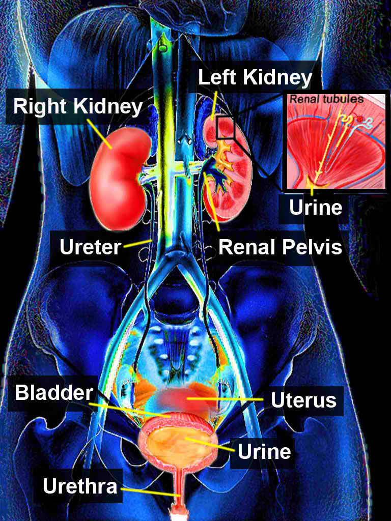
<p>Urinary System. Illustration of the major components making up the urinary system.</p>

Urinary System. Illustration of the major components making up the urinary system.


Contributed by T Silappathikaram

Details

Updated:

7/31/2023 8:27:44 PM

References


[1]

Nasir AA, Ameh EA, Abdur-Rahman LO, Adeniran JO, Abraham MK. Posterior urethral valve. World journal of pediatrics : WJP. 2011 Aug:7(3):205-16. doi: 10.1007/s12519-011-0289-1. Epub 2011 Aug 7     [PubMed PMID: 21822988]


[2]

Dewan PA, Zappala SM, Ransley PG, Duffy PG. Endoscopic reappraisal of the morphology of congenital obstruction of the posterior urethra. British journal of urology. 1992 Oct:70(4):439-44     [PubMed PMID: 1450856]


[3]

Dewan PA, Keenan RJ, Morris LL, Le Quesne GW. Congenital urethral obstruction: Cobb's collar or prolapsed congenital obstructive posterior urethral membrane (COPUM). British journal of urology. 1994 Jan:73(1):91-5     [PubMed PMID: 8298906]


[4]

STEPHENS FD. Urethral obstruction in childhood; the use of urethrography in diagnosis. The Australian and New Zealand journal of surgery. 1955 Nov:25(2):89-109     [PubMed PMID: 13269355]


[5]

Casella DP, Tomaszewski JJ, Ost MC. Posterior urethral valves: renal failure and prenatal treatment. International journal of nephrology. 2012:2012():351067. doi: 10.1155/2012/351067. Epub 2011 Aug 9     [PubMed PMID: 21860792]

Level 3 (low-level) evidence

[6]

Krishnan A, de Souza A, Konijeti R, Baskin LS. The anatomy and embryology of posterior urethral valves. The Journal of urology. 2006 Apr:175(4):1214-20     [PubMed PMID: 16515962]


[7]

Thakkar D, Deshpande AV, Kennedy SE. Epidemiology and demography of recently diagnosed cases of posterior urethral valves. Pediatric research. 2014 Dec:76(6):560-3. doi: 10.1038/pr.2014.134. Epub 2014 Sep 8     [PubMed PMID: 25198372]

Level 3 (low-level) evidence

[8]

Brownlee E, Wragg R, Robb A, Chandran H, Knight M, McCarthy L, BAPS-CASS. Current epidemiology and antenatal presentation of posterior urethral valves: Outcome of BAPS CASS National Audit. Journal of pediatric surgery. 2019 Feb:54(2):318-321. doi: 10.1016/j.jpedsurg.2018.10.091. Epub 2018 Nov 7     [PubMed PMID: 30528204]


[9]

Sarhan OM. Posterior urethral valves: Impact of low birth weight and preterm delivery on the final renal outcome. Arab journal of urology. 2017 Jun:15(2):159-165. doi: 10.1016/j.aju.2017.01.005. Epub 2017 Mar 7     [PubMed PMID: 29071146]


[10]

Hodges SJ, Patel B, McLorie G, Atala A. Posterior urethral valves. TheScientificWorldJournal. 2009 Oct 14:9():1119-26. doi: 10.1100/tsw.2009.127. Epub 2009 Oct 14     [PubMed PMID: 19838598]


[11]

Cohen HL, Zinn HL, Patel A, Zinn DL, Haller JO. Prenatal sonographic diagnosis of posterior urethral valves: identification of valves and thickening of the posterior urethral wall. Journal of clinical ultrasound : JCU. 1998 Sep:26(7):366-70     [PubMed PMID: 9719988]


[12]

Sharma S, Joshi M, Gupta DK, Abraham M, Mathur P, Mahajan JK, Gangopadhyay AN, Rattan SK, Vora R, Prasad GR, Bhattacharya NC, Samuj R, Rao KLN, Basu AK. Consensus on the Management of Posterior Urethral Valves from Antenatal Period to Puberty. Journal of Indian Association of Pediatric Surgeons. 2019 Jan-Mar:24(1):4-14. doi: 10.4103/jiaps.JIAPS_148_18. Epub     [PubMed PMID: 30686881]

Level 3 (low-level) evidence

[13]

Bernardes LS, Aksnes G, Saada J, Masse V, Elie C, Dumez Y, Lortat-Jacob SL, Benachi A. Keyhole sign: how specific is it for the diagnosis of posterior urethral valves? Ultrasound in obstetrics & gynecology : the official journal of the International Society of Ultrasound in Obstetrics and Gynecology. 2009 Oct:34(4):419-23. doi: 10.1002/uog.6413. Epub     [PubMed PMID: 19642115]


[14]

Nicolaides KH, Cheng HH, Snijders RJ, Moniz CF. Fetal urine biochemistry in the assessment of obstructive uropathy. American journal of obstetrics and gynecology. 1992 Mar:166(3):932-7     [PubMed PMID: 1550169]


[15]

Holmes N, Harrison MR, Baskin LS. Fetal surgery for posterior urethral valves: long-term postnatal outcomes. Pediatrics. 2001 Jul:108(1):E7     [PubMed PMID: 11433086]


[16]

Sananes N, Cruz-Martinez R, Favre R, Ordorica-Flores R, Moog R, Zaloszy A, Giron AM, Ruano R. Two-year outcomes after diagnostic and therapeutic fetal cystoscopy for lower urinary tract obstruction. Prenatal diagnosis. 2016 Apr:36(4):297-303. doi: 10.1002/pd.4771. Epub 2016 Feb 17     [PubMed PMID: 26739350]


[17]

Deshpande AV. Current strategies to predict and manage sequelae of posterior urethral valves in children. Pediatric nephrology (Berlin, Germany). 2018 Oct:33(10):1651-1661. doi: 10.1007/s00467-017-3815-0. Epub 2017 Nov 20     [PubMed PMID: 29159472]


[18]

Tourchi A, Kajbafzadeh AM, Aryan Z, Ebadi M. The management of vesicoureteral reflux in the setting of posterior urethral valve with emphasis on bladder function and renal outcome: a single center cohort study. Urology. 2014 Jan:83(1):199-205. doi: 10.1016/j.urology.2013.07.033. Epub 2013 Oct 19     [PubMed PMID: 24149109]


[19]

Sudarsanan B, Nasir AA, Puzhankara R, Kedari PM, Unnithan GR, Damisetti KR. Posterior urethral valves: a single center experience over 7 years. Pediatric surgery international. 2009 Mar:25(3):283-7. doi: 10.1007/s00383-009-2332-z. Epub 2009 Jan 29     [PubMed PMID: 19184051]


[20]

Chua ME, Ming JM, Carter S, El Hout Y, Koyle MA, Noone D, Farhat WA, Lorenzo AJ, Bägli DJ. Impact of Adjuvant Urinary Diversion versus Valve Ablation Alone on Progression from Chronic to End Stage Renal Disease in Posterior Urethral Valves: A Single Institution 15-Year Time-to-Event Analysis. The Journal of urology. 2018 Mar:199(3):824-830. doi: 10.1016/j.juro.2017.10.024. Epub 2017 Oct 20     [PubMed PMID: 29061539]


[21]

Pohl M, Mentzel HJ, Vogt S, Walther M, Rönnefarth G, John U. Risk factors for renal insufficiency in children with urethral valves. Pediatric nephrology (Berlin, Germany). 2012 Mar:27(3):443-50. doi: 10.1007/s00467-011-1999-2. Epub 2011 Oct 19     [PubMed PMID: 22009479]


[22]

DeFoor W, Clark C, Jackson E, Reddy P, Minevich E, Sheldon C. Risk factors for end stage renal disease in children with posterior urethral valves. The Journal of urology. 2008 Oct:180(4 Suppl):1705-8; discussion 1708. doi: 10.1016/j.juro.2008.03.090. Epub 2008 Aug 16     [PubMed PMID: 18708224]


[23]

Sarhan O, Zaccaria I, Macher MA, Muller F, Vuillard E, Delezoide AL, Sebag G, Oury JF, Aigrain Y, El-Ghoneimi A. Long-term outcome of prenatally detected posterior urethral valves: single center study of 65 cases managed by primary valve ablation. The Journal of urology. 2008 Jan:179(1):307-12; discussion 312-3     [PubMed PMID: 18006017]

Level 3 (low-level) evidence

[24]

McLeod DJ, Szymanski KM, Gong E, Granberg C, Reddy P, Sebastião Y, Fuchs M, Gargollo P, Whittam B, VanderBrink BA, Pediatric Urology Midwest Alliance (PUMA). Renal Replacement Therapy and Intermittent Catheterization Risk in Posterior Urethral Valves. Pediatrics. 2019 Mar:143(3):. pii: e20182656. doi: 10.1542/peds.2018-2656. Epub 2019 Feb 1     [PubMed PMID: 30709926]


[25]

Narva AS, Norton JM, Boulware LE. Educating Patients about CKD: The Path to Self-Management and Patient-Centered Care. Clinical journal of the American Society of Nephrology : CJASN. 2016 Apr 7:11(4):694-703. doi: 10.2215/CJN.07680715. Epub 2015 Nov 4     [PubMed PMID: 26536899]


[26]

Crowley R, Wolfe I, Lock K, McKee M. Improving the transition between paediatric and adult healthcare: a systematic review. Archives of disease in childhood. 2011 Jun:96(6):548-53. doi: 10.1136/adc.2010.202473. Epub 2011 Mar 8     [PubMed PMID: 21388969]

Level 1 (high-level) evidence

[27]

Morris RK, Malin GL, Quinlan-Jones E, Middleton LJ, Hemming K, Burke D, Daniels JP, Khan KS, Deeks J, Kilby MD, Percutaneous vesicoamniotic shunting in Lower Urinary Tract Obstruction (PLUTO) Collaborative Group. Percutaneous vesicoamniotic shunting versus conservative management for fetal lower urinary tract obstruction (PLUTO): a randomised trial. Lancet (London, England). 2013 Nov 2:382(9903):1496-506. doi: 10.1016/S0140-6736(13)60992-7. Epub 2013 Aug 14     [PubMed PMID: 23953766]

Level 1 (high-level) evidence