Introduction
The cell is the fundamental organizational unit of life. All living things are composed of cells, which then further subdivide based on the presence or absence of the nucleus, into two types: eukaryotic cells (Greek, Eu=true, karyo=nut, nucleus) - these cells are present in all the human, animal and plants with a clear, distinct nucleus. Prokaryotic cells are some bacteria and blue-green algae which do not contain a clear and distinct nucleus, but the nuclear material is spread within the cytoplasm. The cells with a similar structure and function come together to form tissue. The tissues basically classify as four different types, namely, the epithelial, connective, muscular, and nervous tissues. A combination of these tissues is present in an organ. The total number of cells, their type, size, and shape finally determines the size, structure, and function of each particular organism.[1]
A human body is composed of close to fifty to a hundred trillion cells.[2] They show high diversity in their sizes, structure, number, and function. The human brain itself is estimated to contain around a hundred billion neurons and the same amount of supporting glial cells.[3] The cell size varies significantly with the diameter, ranging from 7.5 µm (RBC) to 150 µm (Ovum). They are classified into different types and intended to perform specialized activities like nerve cells, muscle cells, etc. Classically, estimates are that there are almost 200 different types of cells in an adult human body based on a histological or morphological perspective. Nonetheless, our knowledge of the cells that make up the human body, how they vary from person to person, or throughout development and in health or disease, is still quite limited. Cells have been studied, classified, and precisely characterized since the seventeenth century when Robert Hooke first identified them under the microscope. However, we still have not yet determined all the molecular constituents of cells and how they associate to form tissues and organ systems. Hence, there may be many cell types not known to us, many cellular alterations, and interactions of which we are still unaware. In 2017, a huge undertaking, namely, the International Human Cell Atlas Initiative, came into being to develop a comprehensive reference map of all human cells. This initiative aims to provide a basis for understanding human health in addition to diagnosing and treating diseases.
Understanding the details of each type of cell is a study on its own. Hence, in the present article, only some of the basic and essential features of a normal cell will be described.
Structure
When one studies the cell, each one is surrounded by a fragile plasma membrane that separates the cell and its contents from the surrounding. Each cell has a protoplasm, which is a combination of cytoplasm and the nucleus. The cytoplasm consists of the cytosol (hyaloplasm) and the organelles. The cell membrane acts as a fencing material.
A cell is a combination of different structures:
Cell Organelle:
Just like our bodies need different specialized types of organs and tissues to function properly, the cells also have specialized parts called organelles. Each of these performs a different function. Each organelle has a specific and unique job without which the cell will not be able to survive. A few examples of these being, Golgi bodies, mitochondria, ribosomes, lysosomes, endoplasmic reticulum, etc.
Cell inclusions: These are small non-living structures which are within the cell cytoplasm. A few examples being, melanin, glycogen, enzymes, etc.
The cytosol present in the cytoplasm is watery, but the cytoplasm is gel-like because of the presence of organelles. The cytoplasm consists of hundreds or even thousands of little organs or organelles. The cytosol also has within itself cytoskeleton entirely made up of microfilaments, microtubules, and intermediate filaments. This cytoskeleton gives a definite shape to the cell and also helps in maintaining the same.[4]
Function
Types of cells:
The cells are of different types with a unique function of its own. Estimates are that more than 200 different types of cells are present in an adult human. Some of them are as follows:
- Stem cells: These are the primary cells from which all the other cells arise. These are again of different types for each kind of cell.
- Muscle cells: These are of basically of three types - skeletal, smooth and cardiac cells
- Bone cells: These cells make up the bone. They are of three kinds - osteocytes, osteoclasts, and osteoblasts
- Cartilage cells: These are three types - chondrocytes, chondroblast, and chondroclasts
- Blood cells: They are many types - Platelets, red blood cells, white blood cells, neutrophils, basophils, etc.
- Sex cells: They are two types - sperms and ovum. Further, they pass through different stages of development like (in males) the primordial cells, primary spermatocytes, secondary spermatocytes, spermatids, and finally reach the stage of mature sperms. Also, we find supporting cells like Sertoli cells and hormone-secreting cells like Leydig cells,
- Nervous cells: They are of different types - neuron, oligodendrocytes, astrocytes, microglia, etc.[5]
- Fat cells: These are lipocytes.
General functions of the cells:
As there are many different types of cells, so are their functions. Hence we will summarize some of the essential functions of these into the following.
- Structure: The cells help in giving, and maintaining the specific shape and structure for each part of the body.
- Growth: The stem cells give rise to the development of new types of cells by cell division. There is a spurt in growth in the size/number of the cells during childhood, but get limited when reaching adulthood. This process is regulated by growth-promoting genes that are different for each organ and tissue of the body.[6]
- Apoptosis: It is a process of self-digestion and removal of the dead and worn-out cells.[7] It is necessary for the remodeling, morphogenesis, and regeneration of cells and tissues. It abnormally high in the case of degenerative diseases and too little in cancer cells.[8]
- Metabolism: This takes place by protein, carbohydrate, and lipid synthesis necessary for the cells for growth and their proliferation.[9]
- Regulation of temperature: The metabolism helps in the production of the energy by the mitochondria essential for the function of the cell and body. And with the production of energy, there is an increase in temperature.
- Communication: We always think of neurons playing a significant role in transmitting information from the brain to different parts of the body. But there are also other types of communication between the cells and within by the enzymes, hormones, and others. It is a well-known fact that cells are in continuous interaction with each other for different purposes like metabolism, growth, development, division, transport, etc.[10]
- Transportation of the substances across the membrane: The membrane around the cell show selective permeability. Transport can be active or passive. Some of the elements, like water, carbon dioxide, oxygen, glucose, etc.
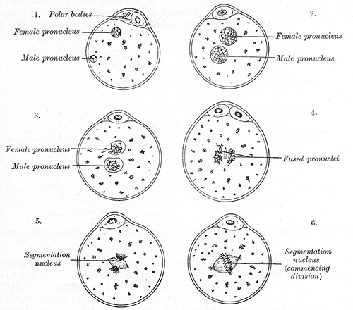
- Reproduction: The cell division and multiplication take place in almost all cells of the body. But when the male and female sex cells come in contact and fuse, it leads to the production of a totally new individual, in the form of a zygote.
The Cell Cycle:
The changes taking place in a cell in a repeated cyclical manner leading to the production of daughter cells is called the cell cycle. Most of the cells of the body divide and replicate, but not all cells show similar patterns of cell cycles. Some cells divided mitotically throughout life like the stem cells, some do divide at an early age and later stop dividing even when damaged like neurons, but some cells divide when there is a demand like the liver cells.
The cell usually divides by two methods:
- Mitosis
- Meiosis
Interphase: It is a state between two mitotic divisions. It further subdivides into the G1 stage, S stage, and G2 stages (G=Gap).
Mitosis takes place in somatic cells of the body. This process is necessary for two reasons: the growth of the organism and the replacement of the dead/worn-out cells. Here the cells divide to produce two daughters that are exact copies of the mother cell.
Meiosis: This kind of cell division takes place in the germ cells - the sperms in the males and the ovum in the females. These germ cells are in the sex gametes - the testis in males and the ovaries in females. In this kind of cell division, the mother cell produces daughter cells, not the copies but unique of its own.
Mitosis (M stage): It subdivides into four stages:
- Prophase
- Metaphase
- Anaphase
- Telophase
Meiosis: It has first and second meiotic divisions.
First Meiotic Division: It is a lengthy division and is divided further into the following four stages
- Leptotene
- Zygotene
- Pachytene
- Diplotene
Second Meiotic Division: It is said to be similar to mitosis except that:
- The DNA content reduces to half.
- The daughter cells are not identical to the mother cell.
Tissue Preparation
The cells are observable in a viable state by growing them in culture suitable for studying them under microscopy. The term for such growth is in vitro (Latin, vitrum=glass). In such an environment, the cells continue to grow with continuous cell lines and perform functions in cohesion to the cells of the body.
Whenever the cells are isolated from their surrounding for the study, they lose their structure, function, and reveal very little of their actual living arrangement in healthy and diseased. Hence their structural integrity is maintained by taking very thin sections along with their surrounding tissue that are suitable to study under light and electron microscope. They are labeled by tracer materials so that their history of sections at different time intervals can be recalled. The sections are so thin and fragile that they require mounting on glass slides for secure handling.
For the light microscopic study, the sections require preparation by the paraffin technique. This preparation needs the following eight chronological steps:
- Tissue sampling: The sample tissue is obtained by surgical excision or biopsy, with the clearing of all connective tissue and undesired structures; this is done by sharp instruments so that the actual structure of the cells/tissue under consideration does not become distorted. The tissue to be studied should not be more than 1 cm. for better fixation.
- Tissue fixation: Once dissected out, the sample requires immediate fixing. Fixation hardens the soft tissue and prevents any postmortem changes and distortion of the cells/tissues. Formalin is the fixator used, usually for light microscopic study. Some of the advantage of fixation apart from preventing postmortem degeneration are better staining of tissue, kill all pathogenic microorganisms (safe to handle), the release of cellular hydrolytic enzymes, etc.
- Dehydration: This occurs by passing the fixed tissue by increasing the strengths of alcohol until it reaches absolute alcohol.
- Clearing: Now, one must remove the alcohol present in the tissue, which takes place by passing the tissue through successive changes of xylol to remove all the alcohol.
- Embedding: Tissue containing the xylol is now passed through successive changes of warm paraffin so that all the spaces in the tissue occupied previously by water and now by xylol becomes replaced with paraffin wax. Soon the tissue hardens on cooling the wax.
- Sectioning: The tissue is now ready for sectioning. Tissue sections are taken with an automated instrument called microtome, which gives fragile sections ranging from 1 to 9 micrometers. Light microscopy usually requires sections of a thickness of 4 to 8 micrometers. If still thinner sections are necessary, then embedding is done in plastic or epoxy resin and not paraffin wax.
- Mounting and staining: The thin sections are mounted on the glass slides and washed with xylol to remove the paraffin wax. Then it passed through decreasing strengths of alcohol and finally washed with water. Now the tissue on the slide is ready for staining. The dewaxed sections are stained with hematoxylin and eosin stains (H&E).[11]
Histochemistry and Cytochemistry
Cytochemistry is the analysis, visualization, and identification of microanatomical locations of biochemical content and its environment, within a cell. This visualization is carried out on histological sections and referred to using electron microscopic techniques or biochemical analyses. Many sophisticated methods are useful for cytochemical analyses such as enzyme cytochemistry, microincineration, microspectrophotometry, radioautography, cryo-techniques, X-ray microanalysis, and immunocytochemistry. These techniques are instrumental in providing discrete information about the ultrastructure and organelles of the cells. Microincineration techniques can provide insight into the distribution of mineral elements, such as calcium (Ca), sodium (Na), potassium (K), etc., in cells, tissues, and organs. Enzymes can be localized within the cell or tissue by of enzymatic conversion of specific chromogenic substrates to give visible results. It is thus a method of cytochemical staining that gives enzymatic colorimetric reactions. Alternatively, microspectrophotometry can measure the spectra of intracellular organelles using electromagnetic radiation of different wavelengths. Organelles and ultrastructures are visualizable by their differential interaction with these wavelengths. Radiography and X-rays help to visualize the pattern of the positioning of radioactively labeled isotopes in cells and tissues. Immunochemical techniques employ the use of specifically labeled antibodies to visualize anatomical structures and their localization based on the specific protein or antigen in cells to the specific primary antibody. Overall, cytochemistry of the cell implies to detection and identification of the biochemical content within a cell. It has helped to elucidate the functional features of cells and also tissue under different pathological, physiological, and experimental conditions.
In contrast, histochemistry is the identification and distribution of chemical components within and between cells. It utilizes a combination of histological and biochemical techniques like stains and indicators and employs light and electron microscopy to study the chemical constituents of the cells and tissues. The technique is imperative to visualize biological structures. Histochemical methods help with understanding the molecular basis of different pathologies and, most specifically, cancer progression. With the advent of modern technology, live-cell staining is also possible. Fluorescent dyes such as intrinsic fluorophores, genetically encoded fluorophores, self-labeling, or ligase based tag systems, have enabled the use of histochemical analyses on live cells. The application of histochemistry in investigating the structure and function of hard tissues such as odontoblasts has provided therapeutic approaches towards dentin mineralization.[12] Immunohistochemistry methods are also useful in elucidating nerve cell differentiation during development.[13] Furthermore, histochemical techniques can function as a method for regenerative and reparative medicine. Histochemical techniques regularly assist in the diagnosis of metabolic disorders and several disease pathologies.
Microscopy, Light
When we study the cell under a light microscope, we can see the structure of the cell and its organelles. Some of the finer details are only appreciated when examined with a more advanced electron microscope.
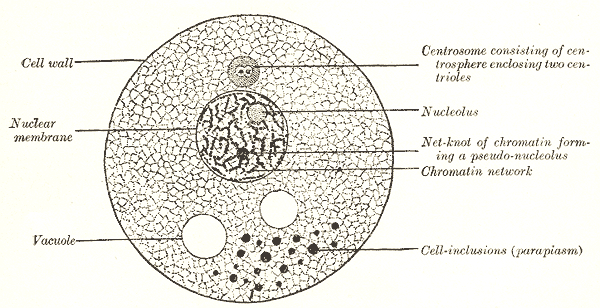
The Cytoplasm (Greek, kytos=covering): Also called plasma (Greek, plasma=molded), as it looks as though it is molded around the nucleus. It is highly crowded with two types of broadly divided components: cytoplasmic organelles and macromolecules. The cytoplasmic organelles (minute organs) will be floating within the cytoplasmic matrix (cytosol). There will also be non-essential structures called inclusion bodies such as pigmented granules or stored fat droplets.[14]
The Nucleus: It is the most rigid and largest cell organelle.[15] It usually occupies the central portion of the cell. Its name derives from the fact that it looks like a nut in the midst of a shell (Latin, nux=nut, Greek, karyo=nut). The nucleus is programmed to control all the functions of the cell. Hence it is referred to as the “brain” of the cell. Surrounding the nucleus is a double-layered thin membrane called nuclear envelope (nuclear membrane) that shows innumerable perforations. These pores are called nuclear pores, which are specialized to have selective permeability.[16] At the center of the nucleus, we find one or more small non-membranous bodies called nucleolus/nucleoli made up of RNA that helps make ribosomes. The nuclei are heterogeneous structures with electron-dense (dark) and electron-lucent (light) areas. These dense areas called heterochromatin consist of tightly coiled inactive chromatin found in irregular clumps often around the periphery of the nucleus. On the other hand, the electron-lucent nuclear material called euchromatin represents part of DNA that is active in RNA synthesis. The heterochromatin and euchromatin are collectively called chromatin (Greek, chroma=colour) as they show affinity towards certain dyes. It contains DNA molecules (hereditary molecules), which appear as granules or threads called chromatin when a cell is in a non-dividing state. When dividing, they look like short, rod-like, tightly coiled structures and now called chromosomes. The human cells typically contain 46 chromosomes (except mature sex cells which contain a haploid number of chromosomes, i.e., 23 chromosomes). The DNA molecules carry the master code for making all of the enzymes and other proteins of a cell. Thus they dictate both the structure and the function of the cells. The non-staining component within which the nucleoli are suspended is known as the nuclear sap.
Ribosomes: The ribosomes are approximately 15 nm in diameter and appear as attached as well as free structures. Many of them attach to the rough endoplasmic reticulum. And many of them are also seen scattered free throughout the cytoplasm. When present alone, they are said to be monosomes and when present in groups, then called polyribosomes. Each of these ribosomes has subunits composed of ribonucleic acid (RNA). The RNA can be rRNA (ribosomal RNA), mRNA (messenger RNA), or tRNA (transfer RNA). Their function is to synthesize proteins for the use, both within and outside the cell. Hence it is said to be the protein factory of a cell. They “translates” the DNA into proteins.
Endoplasmic Reticulum (ER): It means a ‘network’ present towards the center of the cytoplasm and is said to be one of the largest organelles. It is a membrane channel made up of cisterns or tubules. The cytoplasm within these tubules is called vacuoplasm, and that outside is called hyaloplasm or cytosol. It is a complex organelle performing different functions like protein synthesis, calcium storage, steroid synthesis, and lipid metabolism. The ER is said to have different shapes, and each is associated with a specific function. It has been observed that the cells involved in synthesizing excessive amounts of proteins have more sheets, and those involved in lipid synthesis have more tubules.[17] There are two types of ER: the rough and the smooth. The rough endoplasmic reticulum is covered by many ribosomes and helps in protein synthesis. The smooth endoplasmic reticulum synthesizes specific lipids and carbohydrates.
Rough endoplasmic reticulum (rER): They are identified by the membranes with a rough outer surface that is due to attached ribosomes. They act as a miniature circulatory system for the cell or the internal delivery system of the cell. Their lumen is continuous with those of smooth endoplasmic reticulum and perinuclear space. Their primary function is the modification of the synthesize proteins that are used up by the cells.
Smooth endoplasmic reticulum (sER): These structures are membranes having a smooth outer surface, as they are not covered by any ribosomes. Their primary function is to produce the lipids and further processing of membrane proteins that are synthesized by the rough endoplasmic reticulum. They also help in detoxification of drugs.
Typically, only well-folded proteins are delivered to the Golgi apparatus for further needful. If there is any defect or incomplete folding of proteins, they undergo ER-associated degradation. In certain conditions where there is an increase in the production of protein and accumulation of defectively folded proteins, it leads to a condition called ER stress.[18]
Golgi Apparatus: The Golgi apparatus are irregular bodies present near the nucleus of a cell. They are observable under the light microscope by staining with silver salts. When observed under the electron microscope, one can appreciate that a single-layered membrane binds them in a ribbon-like fashion. They are similar to sER and made up of stacks of cisternae and small rounded vesicles at the periphery. They help in protein biosynthesis and packaging protein molecules for export from the cell. The materials from the ER will reach the Golgi bodies in the form of vesicles. From a functional point of view, the Golgi apparatus divides into three regions: cis Golgi, trans Golgi, and medial Golgi.
Lysosomes: They are vesicles that pinch off from the Golgi apparatus. They contain chemicals (enzymes) that help in degrading and recycling cellular waste by a process known as autophagy. This process helps in eliminating the unwanted molecules and foreign particles like the bacteria/virus/foreign bodies that get into the cells. This process occurs by chemicals like lysosomal hydrolases that are released by the lysosomes. Almost 60 different kinds of hydrolases have been identified.[19] Hence they are known as the ‘Digestive Bags’ or ‘Cellular Garbage Disposal units.’ Sometimes they are also called the “suicide bag” because, in some certain rare conditions, the lysosomes can release its chemicals, thus killing the cell itself. Recent research has given a new dimension to the lysosomes by discovering that they are not only dead-end bags but also regulate energy metabolism and cellular clearance. They are also said to play a role in plasma membrane repair, bone resorption, and immune response.[20]
Mitochondria: They are in the form of rods or granules and hence the name mitochondria (mitos=granules, chondrium=rod). They are said to be the ‘Power Plant/house’ of the cell as they will provide all the energy needed by a cell to move, divide, contract, produce secretory products, and all other functions of a cell; this occurs by breaking down the food that helps in making the ATP, which is the principal fuel for all cell activities that require energy. Their size varies from 0.5 to 2 micrometers in length. Their number in each cell depends on the activity of the concerned cell itself being high in a metabolically active cell and low in an inactive one. They have two membranes: inner and outer. The inner layer possesses many folds, and these folds are called cristae. Embedded within the inner membrane is granular material called the matrix, which contains the main enzymes that are essential for the production of adenosine triphosphate (ATP). The location of cellular respiration (the process that makes the cell energy).
Vesicles & Vacuoles: These structures act as the storage compartments of the cell. They usually hold proteins, wastes, food, etc. In the case of plant cells, they hold water within the vacuoles.
Centrioles & Centrosomes: These are essential structures that play in role in the cell division and replication.
The Cell Membrane: It is also known as the plasma membrane or plasmalemma (Greek, lemma=bark). It is a tri-laminar membrane made up predominantly of lipids (fats). It is 7.5 nm in thickness and so thin that it can be seen only by an electron microscope. It also contains a small number of proteins and carbohydrates. These are the lipids like phospholipids, cholesterol, and glycolipids and proteins like integral membrane protein, peripheral membrane protein, and glycoproteins. The different types of proteins help in the active transport of chemicals, food, and wastes. The membrane basically keeps the cell together, separated from the surrounding, gives a definite shape, and maintains the same. In case it breaks, the contents of the cell will spill out. The membrane shows selective permeability being highly permeable to oxygen and water but limited to that of sodium ions, potassium ions, etc. Some of the large molecules enter the cell by endocytosis. They also bear specific receptors for specific enzymes or hormones. Some cells are also specialized to engulf foreign materials by a process called phagocytosis. When engulfing small molecules of fluid, the process is called pinocytosis.
Some of the cells show projection from its surfaces in the form of cilia, flagella, or microvilli.
Pathophysiology
Oxygen, being a bi-radical, is reactive with various metal ions and biological molecules called oxidation. However, the very process of respiration (mitochondrial respiration) produces many forms of reactive oxygen species, such as superoxide anion radical (O2·−), hydrogen peroxide (H2O2), and hydroxyl radical (·OH). All biological molecules, including DNA, are susceptible to this oxidation. However, the body utilizes various antioxidant mechanisms such as superoxide dismutases, peroxidases, peroxiredoxins, and glutathione and glutaredoxins to manage and reduce the amount of oxidation induces stress. DNA undergoes constant damage through its cellular milieu and cellular metabolites, in addition to external mutagens, which may subject DNA to strand breakage during replication. Cyclin-dependent kinases (CDK) that are inherent to the regulation of the cell cycle also play an important role in DNA repair.[21] Double-stranded breaks (DSB) are the most toxic DNA lesions. If not repaired, or incorrectly repaired, they may result in loss of heterozygosity or can generate gross chromosomal rearrangements. Single-stranded breaks, depurination, depyrimidination, O6 methylguanines, and cytosine deamination are similar types of DNA damages. If not corrected by the DNA repair system may lead to the development of pathologies. The repair system that corrects DNA damage includes, nonhomologous DNA end joining (NHEJ), base excision repair (BER), single-strand break repair (SSBR), and homologous recombination (HR) and interstrand cross-link repair (ICL). Disorder of HR and ICL can lead to the development of Fanconi anemia, familial breast cancer, and ovarian cancers. NHEJ insufficiency can lead to the development of severe combined immunodeficiency. Pathologies associated with the disorder of BER and SSBR include hyper IgM syndrome and colorectal carcinomas. Defective SSBR also presents with ataxias. Hence, the DNA damage response pathways that protect genome stability are inherently important to prevent neurodegeneration and malignant transformation of cells, in addition to the normal growth and development, immune development, and neurogenesis.[22]
Based on the external stimuli or environmental demands, the cells undergo several changes within themselves. These changes are physiological as well as pathological, leading to progression towards disease. These changes are generally of the following five types, termed as cellular adaptations:
Hypertrophy: It is a condition in which the muscle cells/fibers gain the muscle mass much excessive to their capacity, with no increase in the number of fibers, an overall increase in the size of the structure. It is best seen in the pregnant uterus and muscles of the bodybuilders. This increase in muscle mass has been attributed to a protein growth factor called insulin-like growth factor 1 (IGF-1).[23]
Hyperplasia: It is a condition where the cells divide rapidly in number leading to an overall increase in the size of the structure. It can be physiological or pathological. The best example of a physiological type is the pregnant uterus. Pathologically it can be benign or malignant. Benign prostatic hyperplasia (BPH) is the best example of benign hyperplasia. The endometrial hyperplasia in the endometrial carcinoma is not infrequent.[24] Endometrial hyperplasia is a pathological state where the endometrial glandular tissue and stroma lining the uterus show severe hyperplastic changes.
Atrophy: It is just the opposite of hypertrophy, wherein the cells start to shrink in size leading to an overall decrease in the size of a tissue or organ. The thymus atrophy after mid-adulthood is a classic example of physiological atrophy. Disuse atrophy is a term used for conditions where atrophy takes place in a specific tissue/organs after prolonged disuse of that particular structure. The atrophy of cells/tissues is due to an overall loss of cell organelles, proteins, and cytoplasm.[25]
Metaplasia: It is a condition of change in cellular identity by a replacement of one type of healthy cells with other healthy cells type in a tissue/organ. An abnormal stimulus induces it.[26] This condition commonly presents in the lower end of the esophagus due to chronic gastroesophageal reflux.
Dysplasia: It is a condition where an abnormal arrangement of cells takes place due to a change in their usual growth behavior.
Clinical Significance
1. Mitochondrial cytopathy syndromes: It is a condition where the mitochondrial DNA is abnormal; this might interfere with the function of mitochondria in particular and the function of the cell in general. It is caused either because of nuclear DNA-mutations or mutation within the maternally inherited form of the mitochondrial genome. Even though the symptoms vary widely, they can present in the way of muscle weakness, high levels of lactic acid, and even degenerative lesions of the brain. Diagnosis is by examining the biopsies taken from the muscle cells under an electron microscope. It reveals para-crystalline inclusions of mitochondria, which is the characteristic feature of this disorder. Investigations also include the measurement of serum lactate, CSF lactate, and some neuroradiological tests. Almost 200 different types of mutations of mitochondrial DNA diseases have been identified, and more are being detected with better facilities and knowledge.[27] Dysregulated or mutated mitochondria have also been linked to malignant changes of the hematopoietic stem cells leading to leukemia.[28]
2. Defects of Golgi apparatus: Alteration in the structure and function of the Golgi complex is a feature in various cardiovascular diseases like heart failure, cardiomegaly, and arrhythmia. The dysfunction of the Golgi bodies is also seen in other conditions but primarily related to cardiovascular diseases as it is responsible for transport, biosynthesis, and distribution of proteins of the cardiovascular system.[29]
3. Cell membrane defects: The cell membrane disorders are seen in red blood cells (RBCs) and usually inherited due to mutations in membrane leading to abnormalities within the RBCs, thus reducing their life span and early removal from circulation. This phenomenon is observable in hereditary conditions like hereditary spherocytosis, stomatocytosis, ovalocytosis, and elliptocytosis.[30] The cell membrane defects also occur in conditions like muscular dystrophies, where there are errors in the proteins located in the cell membranes. These disorders include dystrophinopathy, Bethlem myopathy, merosinopathy, dysferlinopathy, sarcoglycanopathies, and caveolinopathy. The defect within the cell membrane may lead to the influx and efflux of different molecules that might trigger degeneration of the muscle cells.[31] Membrane defects also appear in receptor-related disorders like the Grave's disease, some types of diabetes and obesity, multiple sclerosis; altered lipid state such as multiple sclerosis and muscular dystrophy; defective permeability of the membrane-like cystic fibrosis, bacterial toxins leading condition; some disease related to specific transport alterations and cytoskeleton-membrane defects like Chediak-Higashi disease.[32]
4. Lysosome storage diseases (LSDs): The lysosomes contain more than 50 different types of membrane proteins. It is worthwhile to note that the abnormality or dysfunction of some of the specific proteins in lysosomes has led to the detection of almost 50 different types of lysosomal storage diseases. Lysosomal mutation and lysosomal genes predispose the human to a whole array of conditions. The most prevalent diseases with LSDs like obesity, infection, cancer, and neurodegenerative diseases like Parkinson disease, Gaucher disease, and Alzheimer disease.[20]
5. Nuclear defects: Mutation in the nuclear structure and composition are also present in diseases like muscular dystrophy, cancer, aging, cardiomyopathies.[33] Neurodegenerative diseases show abnormal accumulation of pathogenic intranuclear protein aggregation as intranuclear inclusion bodies.[34]
6. Endoplasmic reticulum defects: Researchers have observed that different ER-shaping proteins play a role in diseases such as hereditary spastic paraplegia and Alzheimer disease.[17] There is evidence to prove that prolonged ER stress leads to many diseases like type 2 diabetes, neurodegeneration, liver disease, atherosclerosis, and cancers.[18]
7. Cytoplasmic errors: As we know, every constitution of the cell should be an ideal quantity. Research has observed that an abnormally large amount of cytoplasm leads to defect in the chromosome alignment, spindle pole morphology, and checkpoint signaling leading to chromosome segregation errors.[35]
8. Mutation: Abnormalities in mitosis results from prolonged exposure to radiation (especially nuclear radiation). It can also happen with certain chemicals and drugs. But some cells do not undergo mitosis like a neural cell, cardiac cells. They are said to be in the Go stage.
9. Tumors: The rate of division of cells dramatically varies in different cells. It is highest in the epithelial cells exposed to friction. Rate correlates with demand. Excessive uncontrolled growth leads to a condition commonly known as tumors.[36]