Juvenile Glaucoma

Earn CME/CE in your profession:


Continuing Education Activity

Juvenile open-angle glaucoma is an uncommon and more severe form of primary open-angle glaucoma. It occurs as a result of abnormal development of trabecular meshwork. It is manageable medically or surgically. This activity discusses the etiology and the pathophysiology of the Ashman phenomenon and highlights the role of the interprofessional team in evaluating and treating patients with this condition.

Objectives:

  • Review the etiology for developing juvenile open-angle glaucoma.
  • Outline the typical presentation of a patient with juvenile open-angle glaucoma.
  • Identify the most common complications associated with juvenile open-angle glaucoma.
  • Summarize the importance of improving communication and care coordination amongst the interprofessional team to enhance the delivery of care for patients with juvenile open-angle glaucoma.

Introduction

Glaucoma is a neurodegenerative disorder characterized by the progressive degeneration of the optic disc and visual field loss, for which intraocular pressure (IOP) is the chief modifiable risk factor. Glaucoma is the most common etiology of irreversible blindness. It affects more than 2 million individuals in the United States and 67 million individuals worldwide.

Glaucoma has two main subtypes: open-angle glaucoma and angle-closure glaucoma. Juvenile open-angle glaucoma (JOAG) is an uncommon form of primary open-angle glaucoma, with earlier onset (3 to 40 years of age), higher IOP, and more severe visual field loss compared with adult-onset primary open-angle glaucoma (POAG).

Many studies report that this form of glaucoma typically demonstrates an autosomal dominant inheritance pattern. The myocilin (MYOC) gene is identified abundantly through linkage analysis in the trabecular meshwork (TM) of the affected patients.[1][2][3][4][5][6][7] 

Anatomy of the Eye

The eye can subdivide into two parts: anterior and posterior. The anterior part is located in front of the lens and divides into anterior and posterior chambers. In the posterior chamber, the ciliary body of the eye produces a fluid called aqueous humor. This fluid flows from the posterior chamber into the anterior chamber through the pupil and exits the eye via two pathways: the trabecular and uveoscleral pathways. The majority of aqueous humor outflow flows through the trabecular meshwork (TM) and the canal of Schlemm (SC), which are parts of the trabecular pathway. In the uveoscleral pathway, aqueous humor passes into the supraciliary and suprachoroidal spaces through the ciliary muscle. Finally, both routes drain the aqueous humor into the venous circulation.[8]

Etiology

Juvenile open-angle glaucoma often occurs as a result of abnormal development of trabecular meshwork.[5][7] Therefore, drainage of aqueous humor may not be adequate, leading to increased intraocular pressure and damage to the optic nerve.[8]

Juvenile-onset open-angle glaucoma has many risk factors such as:

  • Male sex: in a study of 125 eyes with JOAG, 64% were male.[5]
  • Myopia: Researchers found 87% of JOAG individuals to be myopic in a study of 23 affected patients.[9]
  • Severe elevation of intraocular pressure IOP: it is a typical feature of juvenile-onset primary open-angle glaucoma. Generally, intraocular pressures range over 40 mmHg.[10]
  • African ancestry[2]
  • MYOC mutation[4]

Epidemiology

The prevalence of juvenile open-angle glaucoma is 0.38 per 100,000, ages ranging between 4 and 20 years.[5] Based on a study from the Dallas Glaucoma Registry, which comprised 376 eyes of 239 childhood glaucoma patients, 4% of patients had juvenile open-angle glaucoma.[11]

Pathophysiology

The pathophysiology of glaucoma remains incompletely understood.[12] However, it appears that the elevated intraocular pressure IOP in juvenile-onset primary open-angle glaucoma patients is due to goniodysgenesis, a failure of anterior chamber development, which includes an immature trabecular meshwork and thickened trabecular beams. Developmental abnormalities of the anterior chamber angle are less apparent with older subjects. Later age presentation of glaucoma may have less severe abnormalities of the trabecular meshwork and tends to involve only one eye.[6]

History and Physical

Juvenile open-angle glaucoma and adult-onset primary open-angle glaucoma have similar findings. A significant number of individuals with juvenile open-angle glaucoma are asymptomatic. Symptoms that may present include an enlarged eye, blurred vision, eye pain, decreased visual acuity, tearing, blinking, and glare. A family history of glaucoma should be taken in addition to other histories to rule out secondary glaucoma.[5][13]

Evaluation

Evaluation of juvenile open-angle glaucoma should include the measurement of IOP and assessment of visual acuity, the optic disc, and the anterior chamber angle.

  • Intraocular pressure: Goldmann Applanation Tonometry is the gold-standard method used to obtain an accurate IOP.[14]
  • Optic disc: optic discs of juvenile open-angle glaucoma tend to be more extensive than adult-onset primary open-angle glaucoma discs.[15]
  • Pachymetry: This is used to measure the thickness of the eye's cornea and is recommended as a part of the pre-operative evaluation of juvenile open-angle glaucoma. Thinner corneas are a risk factor for JOAG.[16]
  • Anterior chamber angle: Will typically appear normal on gonioscopy in patients with juvenile open-angle glaucoma. However, dysgenesis of the angle may be present with severe elevation of intraocular pressure.[17]

Treatment / Management

Treatment for juvenile open-angle glaucoma aims to stop further degeneration of the optic nerve by decreasing the IOP. Imaging of the optic nerve is often necessary to assess disease progression. Frequently, the IOP in juvenile open-angle glaucoma tends to be resistant to medical therapy, and surgical treatment is often needed.[18]

Medical Therapy

The first-line treatment of juvenile open-angle glaucoma is medical therapy, followed by surgery if medical therapy fails. Often medical therapy is applied as a bridge to surgical intervention. According to one study performed on 23 patients, 83% of patients with juvenile glaucoma require filtration surgery (trabeculectomy).[9]

Medical therapy for juvenile open-angle glaucoma includes β blockers, topical carbonic anhydrase inhibitors, prostaglandin analogs, and α-adrenergic agonists. If no contraindications such as asthma exist, β-blockers are often the first-line therapy for juvenile open-angle glaucoma. Topical carbonic anhydrase inhibitors are less effective than β blockers, but they are useful as additional therapy or as alternatives when β blockers are contraindicated. Prostaglandins are more effective in adult glaucoma than childhood glaucoma. The α-adrenergic agonists can be effective but have potential systemic side effects, which request close observation when used in young patients.[19] 

Surgery

Surgical therapy has multiple options, such as trabeculectomy, drainage implants, angle procedures (goniotomy, trabeculotomy), and cycloablative procedures.[20]

For juvenile open-angle glaucoma, trabeculectomy is the backbone of surgical therapy, which has a good outcome. In an Indian study that included 60 eyes of 41 patients, 50 to 87% of cases have reported IOP control without medications for three years after the operation.[21][22][23] Mitomycin C (MMC) or other types of antifibrotics have been used to prevent fibrosis during operation. The addition of mitomycin C (MMC) to trabeculectomy has resulted in lower intraocular pressures. However, there is a higher risk of developing hypotony maculopathy and bleb-related infection.[23]

Glaucoma drainage implants (GDIs) are an alternative surgical option for JOAG; they include many types of implants such as the Ahmed glaucoma valve, Baerveldt implant, and Molteno implant. Glaucoma drainage implants (GDIs) are preferable in the case of conjunctival scarring that prevents trabeculectomy.[24] One retrospective study showed that the Ahmed glaucoma valve has a 100% success rate at one year when used for pediatric glaucoma. The final intraocular pressure IOP was between 6-18 mmHg, and there were no patients that lost vision completely.[25] In a prospective study of 52 eyes with juvenile glaucoma, the Molteno implant resulted in intraocular pressure (IOP) of less than 21 mmHg.[26] Although there are reports showing successful angle procedures in juvenile open-angle glaucoma, they are usually used in congenital glaucoma management instead.[16] 

Laser

Laser trabeculoplasty is an alternative option to treat glaucoma. It works to lower intraocular pressure IOP. Selective laser trabeculoplasty (SLT) is one of the modalities of laser trabeculoplasty, which uses a Q-switched, frequency-doubled Nd:YAG (532 nm) laser. It enhances aqueous outflow through the trabecular meshwork to decrease IOP. SLT induces less coagulative damage and structural change than older laser trabeculoplasty techniques. SLT does not typically provide the same IOP lowering effect as surgical therapy, and due to goniodysgenesis in juvenile open-angle glaucoma, it often is not as effective as other forms of primary open-angle glaucoma.[27]

Differential Diagnosis

Other conditions can also be mistaken for juvenile open-angle glaucoma juvenile open-angle glaucoma as they may present similarly such as:

  • Congenital glaucoma
  • Steroid-induced glaucoma
  • Traumatic glaucoma
  • Inflammatory glaucoma
  • Other forms of primary open-angle glaucoma

Prognosis

The prognosis for juvenile open-angle glaucoma patients is generally favorable if diagnosed and treated early in the disease course. In a Japanese study of 47 eyes with juvenile glaucoma, 21 eyes (45%) had normal visual fields, whereas 13 eyes (28%) were at an advanced stage, most of which were ascribed to a delay in disease diagnosis.[20] Even if the patient needs a trabeculectomy, the patient's IOP can be decreased and visual field loss halted.[28]

Complications

If glaucoma remains untreated, visual impairment and blindness may develop. It is not uncommon for the diagnosis of glaucoma to be delayed since it may be asymptomatic until an advanced stage.[5][29]

Other complications depend on the type of surgery performed. They include:

  • Eye redness or pain
  • Irritation of the cornea
  • Persistent high intraocular pressure
  • Hypotony
  • Choroidal effusions
  • Suprachoroidal hemorrhages
  • Infection
  • Cataract development or progression

Deterrence and Patient Education

Although juvenile open-angle glaucoma is uncommon, patients and parents should receive education about this condition. Since these patients are at an increased risk of developing visual impairment and blindness, they should understand the nature of the disease and its symptoms. Early diagnosis and treatment increase the chance of preserving vision and preventing permanent visual field loss.

Enhancing Healthcare Team Outcomes

Patients with juvenile open-angle glaucoma may initially present to the emergency department physician, primary care provider, or other medical providers. Good communication between health care providers is necessary to improve the patient's vision outcomes. These patients should receive a referral to an ophthalmologist for definitive diagnosis and treatment planning. The prognosis depends on the time of diagnosis and the treatment received. Practice patterns should be developed and followed so that this disease can be recognized early, preventing irreversible loss of vision.[30][31] [Level 5]


(Click Image to Enlarge)
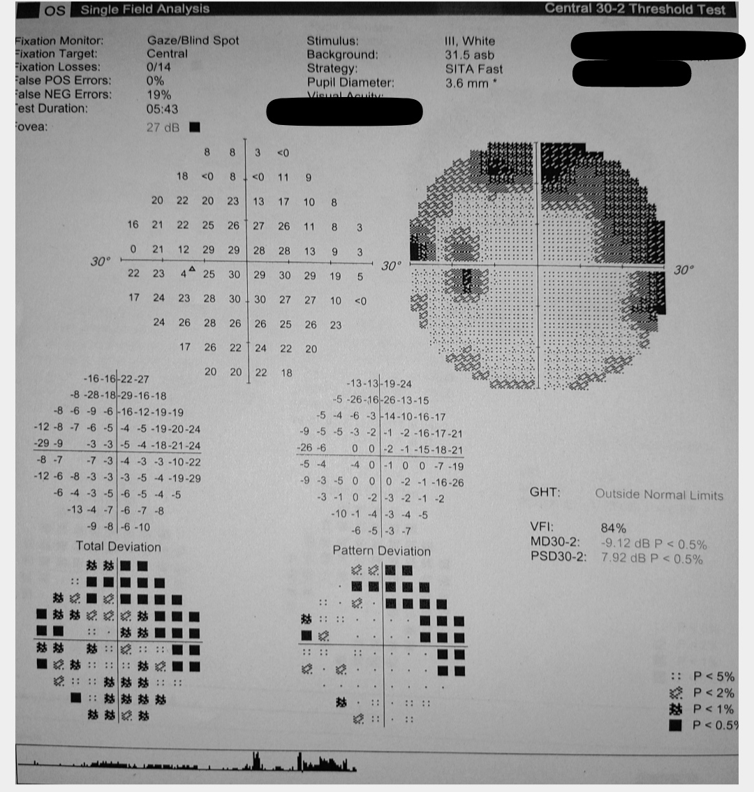
<p>Glaucomatous Visual Field Changes in the Left Eye

Glaucomatous Visual Field Changes in the Left Eye. This image displays the results of a visual field test indicating characteristic changes associated with glaucoma in the left eye, as observed on perimetry.


Contributed by S Shah, MBBS


(Click Image to Enlarge)
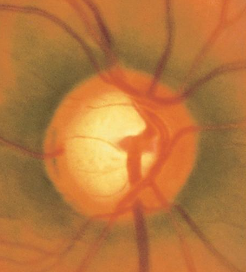
<p>Optic Nerve Cup-to-Disc Ratio of 0

Optic Nerve Cup-to-Disc Ratio of 0.75. A cup-to-disc ratio greater than 0.5 is often associated with glaucoma, and initial loss is typically observed in the inferotemporal and superotemporal poles of the optic disc.


Contributed by AS Huang, MD

Details

Editor:

Kyle Blair

Updated:

12/26/2022 9:31:27 PM

References


[1]

Mantravadi AV,Vadhar N, Glaucoma. Primary care. 2015 Sep;     [PubMed PMID: 26319348]


[2]

Gupta V,Somarajan BI,Gupta S,Chaurasia AK,Kumar S,Dutta P,Gupta V,Sharma A,Tayo BO,Nischal K, The inheritance of juvenile onset primary open angle glaucoma. Clinical genetics. 2017 Aug;     [PubMed PMID: 27779752]


[3]

Arora S,Maeda M,Francis B,Maeda M,Sit AJ,Mosaed S,Nazarali S,Damji KF, Efficacy and safety of ab interno trabeculectomy in juvenile open-angle glaucoma. Canadian journal of ophthalmology. Journal canadien d'ophtalmologie. 2018 Oct;     [PubMed PMID: 30340716]


[4]

Svidnicki PV,Braghini CA,Costa VP,Schimiti RB,de Vasconcellos JPC,de Melo MB, Occurrence of MYOC and CYP1B1 variants in juvenile open angle glaucoma Brazilian patients. Ophthalmic genetics. 2018 Dec;     [PubMed PMID: 30484747]


[5]

Kwun Y,Lee EJ,Han JC,Kee C, Clinical Characteristics of Juvenile-onset Open Angle Glaucoma. Korean journal of ophthalmology : KJO. 2016 Apr;     [PubMed PMID: 27051261]


[6]

Gupta V,Gupta S,Dhawan M,Sharma A,Kapoor KS,Sihota R, Extent of asymmetry and unilaterality among juvenile onset primary open angle glaucoma patients. Clinical     [PubMed PMID: 21631667]


[7]

Goldwyn R,Waltman SR,Becker B, Primary open-angle glaucoma in adolescents and young adults. Archives of ophthalmology (Chicago, Ill. : 1960). 1970 Nov;     [PubMed PMID: 5478882]


[8]

Marshall LL,Hayslett RL,Stevens GA, Therapy for Open-Angle Glaucoma. The Consultant pharmacist : the journal of the American Society of Consultant Pharmacists. 2018 Aug 1;     [PubMed PMID: 30068436]


[9]

Wiggs JL,Del Bono EA,Schuman JS,Hutchinson BT,Walton DS, Clinical features of five pedigrees genetically linked to the juvenile glaucoma locus on chromosome 1q21-q31. Ophthalmology. 1995 Dec;     [PubMed PMID: 9098278]


[10]

Alward WL,Fingert JH,Coote MA,Johnson AT,Lerner SF,Junqua D,Durcan FJ,McCartney PJ,Mackey DA,Sheffield VC,Stone EM, Clinical features associated with mutations in the chromosome 1 open-angle glaucoma gene (GLC1A) The New England journal of medicine. 1998 Apr 9;     [PubMed PMID: 9535666]


[11]

Fung DS,Roensch MA,Kooner KS,Cavanagh HD,Whitson JT, Epidemiology and characteristics of childhood glaucoma: results from the Dallas Glaucoma Registry. Clinical ophthalmology (Auckland, N.Z.). 2013;     [PubMed PMID: 24039394]


[12]

Weinreb RN,Leung CK,Crowston JG,Medeiros FA,Friedman DS,Wiggs JL,Martin KR, Primary open-angle glaucoma. Nature reviews. Disease primers. 2016 Sep 22;     [PubMed PMID: 27654570]


[13]

Geyer O,Mathalone N,Wolf A,Melamud A, [CHILDHOOD GLAUCOMA]. Harefuah. 2019 Feb;     [PubMed PMID: 30779490]


[14]

Bradfield YS,Kaminski BM,Repka MX,Melia M,Davitt BV,Johnson DA,Kraker RT,Manny RE,Matta NS,Schloff S,Weise KK, Comparison of Tono-Pen and Goldmann applanation tonometers for measurement of intraocular pressure in healthy children. Journal of AAPOS : the official publication of the American Association for Pediatric Ophthalmology and Strabismus. 2012 Jun;     [PubMed PMID: 22459105]


[15]

Gupta V,James MK,Singh A,Kumar S,Gupta S,Sharma A,Sihota R,Kennedy DJ, Differences in Optic Disc Characteristics of Primary Congenital Glaucoma, Juvenile, and Adult Onset Open Angle Glaucoma Patients. Journal of glaucoma. 2016 Mar;     [PubMed PMID: 25265002]


[16]

Stangos AN,Whatham AR,Sunaric-Megevand G, Primary viscocanalostomy for juvenile open-angle glaucoma. American journal of ophthalmology. 2005 Sep;     [PubMed PMID: 16084786]


[17]

Gupta V,Srivastava RM,Rao A,Mittal M,Fingert J, Clinical correlates to the goniodysgensis among juvenile-onset primary open-angle glaucoma patients. Graefe's archive for clinical and experimental ophthalmology = Albrecht von Graefes Archiv fur klinische und experimentelle Ophthalmologie. 2013 Jun;     [PubMed PMID: 23358655]


[18]

Johnson AT,Drack AV,Kwitek AE,Cannon RL,Stone EM,Alward WL, Clinical features and linkage analysis of a family with autosomal dominant juvenile glaucoma. Ophthalmology. 1993 Apr;     [PubMed PMID: 8479711]


[19]

Talbot AW,Russell-Eggitt I, Pharmaceutical management of the childhood glaucomas. Expert opinion on pharmacotherapy. 2000 May;     [PubMed PMID: 11249511]

Level 3 (low-level) evidence

[20]

Ikeda H,Ishigooka H,Muto T,Tanihara H,Nagata M, Long-term outcome of trabeculotomy for the treatment of developmental glaucoma. Archives of ophthalmology (Chicago, Ill. : 1960). 2004 Aug;     [PubMed PMID: 15302651]


[21]

Gupta V,Ghosh S,Sujeeth M,Chaudhary S,Gupta S,Chaurasia AK,Sihota R,Gupta A,Kapoor KS, Selective laser trabeculoplasty for primary open-angle glaucoma patients younger than 40 years. Canadian journal of ophthalmology. Journal canadien d'ophtalmologie. 2018 Feb;     [PubMed PMID: 29426447]


[22]

Pathania D,Senthil S,Rao HL,Mandal AK,Garudadari CS, Outcomes of trabeculectomy in juvenile open angle glaucoma. Indian journal of ophthalmology. 2014 Feb;     [PubMed PMID: 23571250]


[23]

Tsai JC,Chang HW,Kao CN,Lai IC,Teng MC, Trabeculectomy with mitomycin C versus trabeculectomy alone for juvenile primary open-angle glaucoma. Ophthalmologica. Journal international d'ophtalmologie. International journal of ophthalmology. Zeitschrift fur Augenheilkunde. 2003 Jan-Feb;     [PubMed PMID: 12566869]


[24]

Ishida K,Mandal AK,Netland PA, Glaucoma drainage implants in pediatric patients. Ophthalmology clinics of North America. 2005 Sep;     [PubMed PMID: 16055000]


[25]

Balekudaru S,Vadalkar J,George R,Vijaya L, The use of Ahmed glaucoma valve in the management of pediatric glaucoma. Journal of AAPOS : the official publication of the American Association for Pediatric Ophthalmology and Strabismus. 2014 Aug;     [PubMed PMID: 25173898]


[26]

Ah-Chan JJ,Molteno AC,Bevin TH,Herbison P, Otago Glaucoma Surgery Outcome Study: follow-up of young patients who underwent Molteno implant surgery. Ophthalmology. 2005 Dec;     [PubMed PMID: 16325709]


[27]

Lamoureux EL,Mcintosh R,Constantinou M,Fenwick EK,Xie J,Casson R,Finkelstein E,Goldberg I,Healey P,Thomas R,Ang GS,Pesudovs K,Crowston J, Comparing the effectiveness of selective laser trabeculoplasty with topical medication as initial treatment (the Glaucoma Initial Treatment Study): study protocol for a randomised controlled trial. Trials. 2015 Sep 11;     [PubMed PMID: 26362541]

Level 1 (high-level) evidence

[28]

Koraszewska-Matuszewska B,Samochowiec-Donocik E,Filipek E, [Prognosis in juvenile glaucoma after trabeculectomy]. Klinika oczna. 2002     [PubMed PMID: 12174451]


[29]

Weinreb RN,Aung T,Medeiros FA, The pathophysiology and treatment of glaucoma: a review. JAMA. 2014 May 14;     [PubMed PMID: 24825645]


[30]

Bouhenni RA,Ricker I,Hertle RW, Prevalence and Clinical Characteristics of Childhood Glaucoma at a Tertiary Care Children's Hospital. Journal of glaucoma. 2019 Jul     [PubMed PMID: 30950965]


[31]

Gupta V,Ganesan VL,Kumar S,Chaurasia AK,Malhotra S,Gupta S, Visual Disability Among Juvenile Open-angle Glaucoma Patients. Journal of glaucoma. 2018 Apr     [PubMed PMID: 29394204]